. 2009 Jun 26;284(26):17634-40.
doi: 10.1074/jbc.M109.008938. Epub 2009 Apr 17.

Generation of induced pluripotent stem cell lines from Tibetan miniature pig

Affiliations

Generation of induced pluripotent stem cell lines from Tibetan miniature pig

Miguel A Esteban et al. J Biol Chem. .

Abstract

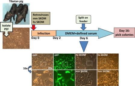
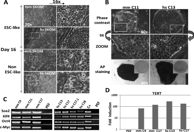
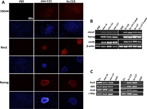
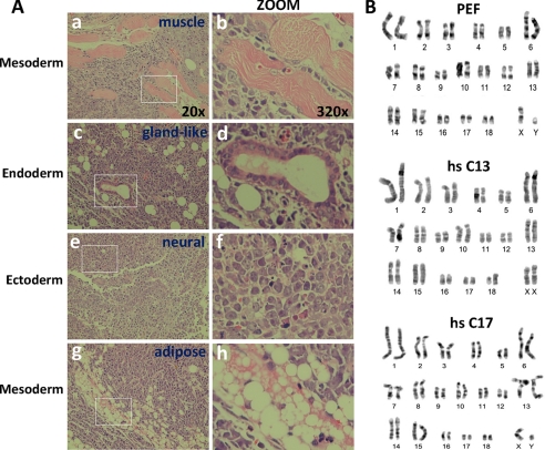
Induced pluripotent stem cell (iPS) technology appears to be a general strategy to generate pluripotent stem cells from any given mammalian species. So far, iPS cells have been reported for mouse, human, rat, and monkey. These four species have also established embryonic stem cell (ESC) lines that serve as the gold standard for pluripotency comparisons. Attempts have been made to generate porcine ESC by various means without success. Here we report the successful generation of pluripotent stem cells from fibroblasts isolated from the Tibetan miniature pig using a modified iPS protocol. The resulting iPS cell lines more closely resemble human ESC than cells from other species, have normal karyotype, stain positive for alkaline phosphatase, express high levels of ESC-like markers (Nanog, Rex1, Lin28, and SSEA4), and can differentiate into teratomas composed of the three germ layers. Because porcine physiology closely resembles human, the iPS cells reported here provide an attractive model to study certain human diseases or assess therapeutic applications of iPS in a large animal model.


Figures

FIGURE 1.
FIGURE 1.
Scheme depicting the generation of porcine iPS cell lines from the moment of viral transduction to colony picking. Captures of Tibetan pigs (5 days old), similar to the strain used for our experiments, are shown on the left. PEF (capture shown on the left) were isolated as described under “Experimental Procedures” and infected with retroviruses coding mouse or human factors. At the bottom of the scheme, captures at day 6 post-infection of PEF transduced with either GFP control retroviruses or SKOM mixtures and culture with defined medium are shown. GFP detected with a fluorescence filter showed almost 100% infection efficiency. Notice the early classical (similar to mouse or human iPS generation) morphology changes (cells becoming rounded and aggregating) only in the SKOM-infected pools. DMEM, Dulbecco's modified Eagle's medium. hs, Homo sapiens; mm, Mus musculus.
FIGURE 2.
FIGURE 2.
Isolation of porcine iPS cell lines. A, colonies with ESC-like characteristics were picked at day 16. Colonies with a variable non-ESC-like morphology were abundant. Magnifications are indicated. B, expanded pig iPS colonies (passage 10 is shown) maintained the original morphology and stained positive for AP after repeated passages. AP staining on 3-cm dishes is shown in reduced size. C, semiquantitative RT-PCR with specific primers shows integration of exogenous mouse or human factors into the genomic DNA of pig iPS clones. Positive (C+) control corresponding to pMX-Sox2 plasmid and negative (uninfected PEF) were included. D, real-time RT-PCR for the reverse telomerase-reverse transcriptase gene shows high expression in selected PEF iPS clones compared with uninfected PEF. Values were normalized with 18S. Pig iPS colonies at passages 9 (mouse SKOM) and 15 (human SKOM) were used. mm, M. musculus; hs, H. sapiens.
FIGURE 3.
FIGURE 3.
Characterization of porcine iPS cell lines. A, immunofluorescence microscopy shows activation of the endogenous ESC program. PEF are shown on the left and stained negative for all markers. Pig iPS colonies at passage 10 were used, note that feeder layers stained negative and serve as an internal comparison. Magnifications are indicated. B, semiquantitative RT-PCR of selected PEF iPS cell lines. eSox2 indicates the endogenous gene; Nanog and Lin28 are also included and β-actin was used as loading control. Uninfected PEF were used as negative control. Treatment of human factors C13 and C17 iPS clones with 5-azacytidine did not further increase expression of ESC markers (right panel). Pig iPS colonies at passages 9 (mouse SKOM) and 15 (human SKOM) were used. C, semiquantitative RT-PCR with primers that specifically amplify the mRNA product of the integrated transgenes shows no silencing in selected iPS cell lines compared with mRNA extracted from infected control cells at day 8 after viral transduction. Water was used as negative control for the PCR. mm, M. musculus; hs, H. sapiens.
FIGURE 4.
FIGURE 4.
Pig iPS cell lines are pluripotent. A, teratoma formation in immunodeficient mice demonstrates differentiation into the three germ layers. Results from the hs SKOM C13 clone (passage 16) are displayed, a similar pattern was observed with hs SKOM C17 (passage 16) (not shown). Magnified pictures are shown on the right, and magnification is indicated for both panels. Mesoderm-derived muscle and fat are shown in a-b and g-h, respectively, a gland-like structure (endoderm derived) in c-d, and neural-like tissue is shown in e-f. B, karyotype analysis demonstrates an equal number of chromosomes (19 pairs) in two different human SKOM iPS cell lines (passage 18) compared with control PEF. Note that the two iPS cell lines correspond to different genders. hs, H. sapiens.

References

    1. Tweedell K. S. ( 2008) Curr. Stem Cell Res. Ther. 3, 151– 162 - PubMed
    1. Pei D. ( 2008) Cell Res. 18, 221– 223 - PubMed
    1. Takahashi K., Yamanaka S. ( 2006) Cell 126, 663– 676 - PubMed
    1. Okita K., Ichisaka T., Yamanaka S. ( 2007) Nature 448, 313– 317 - PubMed
    1. Takahashi K., Tanabe K., Ohnuki M., Narita M., Ichisaka T., Tomoda K., Yamanaka S. ( 2007) Cell 131, 861– 872 - PubMed

Publication types

MeSH terms