. 2009 Jun 19;284(25):16956-16965.
doi: 10.1074/jbc.M806855200. Epub 2009 Apr 16.

Agrin regulation of alpha3 sodium-potassium ATPase activity modulates cardiac myocyte contraction

Affiliations

Agrin regulation of alpha3 sodium-potassium ATPase activity modulates cardiac myocyte contraction

Lutz G W Hilgenberg et al. J Biol Chem. .

Abstract

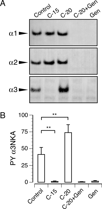
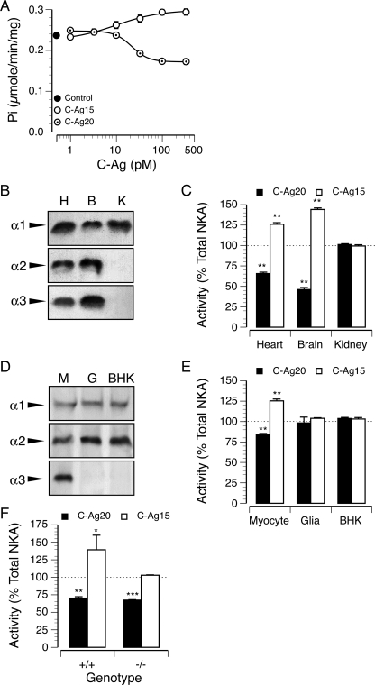
Drugs that inhibit Na,K-ATPases, such as digoxin and ouabain, alter cardiac myocyte contractility. We recently demonstrated that agrin, a protein first identified at the vertebrate neuromuscular junction, binds to and regulates the activity of alpha3 subunit-containing isoforms of the Na,K-ATPase in the mammalian brain. Both agrin and the alpha3 Na,K-ATPase are expressed in heart, but their potential for interaction and effect on cardiac myocyte function was unknown. Here we show that agrin binds to the alpha3 subunit of the Na,K-ATPase in cardiac myocyte membranes, inducing tyrosine phosphorylation and inhibiting activity of the pump. Agrin also triggers a rapid increase in cytoplasmic Na(+) in cardiac myocytes, suggesting a role in cardiac myocyte function. Consistent with this hypothesis, spontaneous contraction frequencies of cultured cardiac myocytes prepared from mice in which agrin expression is blocked by mutation of the Agrn gene are significantly higher than in the wild type. The Agrn mutant phenotype is rescued by acute treatment with recombinant agrin. Furthermore, exposure of wild type myocytes to an agrin antagonist phenocopies the Agrn mutation. These data demonstrate that the basal frequency of myocyte contraction depends on endogenous agrin-alpha3 Na,K-ATPase interaction and suggest that agrin modulation of the alpha3 Na,K-ATPase is important in regulating heart function.


Figures

FIGURE 1.
FIGURE 1.
Agrin binds specifically to the α3 Na,K-ATPase in heart. A and A′, low power photomicrographs of a frozen section through an E18 mouse heart ventricle double labeled for agrin (A) and α3 Na,K-ATPase (A′). Both agrin and the α3 Na,K-ATPase are expressed throughout the ventricular myocardium. Higher levels of α3 Na,K-ATPase can be seen in a subpopulation of cells, possibly developing Purkinje fibers, concentrated in the ventricular septum (arrows) and scattered throughout the myocardium. B and B′, agrin (B) and α3 Na,K-ATPase (B′) are also expressed in adult cardiac muscle fibers. Viewed at higher magnification, agrin appears relatively evenly distributed, whereas α3 Na,K-ATPase is characterized by the presence of more intensely labeled puncta set against a low background. C, typical Western blots (50 μg/protein/lane) of embryo and adult ventricular muscle probed with antibodies against the α3, α2, or α1 subunit of the Na,K-ATPase. The α3 subunit migrates as a single band of a 110 kDa in saline (S)-treated control tissue and is more abundant in embryos than in adults. Cross-linking saline-treated tissue with BS3 generates a ≥300-kDa agrin-α3 subunit complex in which formation is blocked by the presence of either C-Ag15 or C-Ag20, resulting instead in 125- and 130-kDa bands, respectively. In contrast, Western blots of aliquots of the BS3 cross-linked tissue probed with antibodies to α2 or α1 contain only a single band of 110 kDa, evidence that agrin binds specifically to the α3 Na,K-ATPase. Whereas the level of α2 subunit expression increases during development, the α1 subunit is unchanged, confirming similar loading across lanes. D, tissue samples, treated as in C, were solubilized in detergent-containing buffer and immunoprecipitated (IP) with either the anti-agrin serum (Ag) or anti-α3 subunit monoclonal antibody (α3), and the immunoprecipitates were analyzed by Western blotting with the anti-agrin serum. Native agrin glycoprotein immunoprecipitated by the anti-agrin serum from control tissue treated with saline alone is shown for comparison and appears as a broad band of ≥200 kDa. Consistent with interaction between endogenous agrin and the α3 subunit, cross-linking results in the appearance of a high molecular mass species of ≥300 kDa recognized by both the anti-α3 and anti-agrin antibodies. Formation of the agrin-α3 complex is blocked by cross-linking in the presence of a saturating concentration of either C-Ag15 or C-Ag20.
FIGURE 2.
FIGURE 2.
Agrin-dependent tyrosine phosphorylation of the α3 subunit of the Na,K-ATPase. A, typical Western blots show α1, α2, and α3 Na,K-ATPase subunits immunoprecipitated from adult ventricular muscle by an anti-phosphotyrosine antibody. C-Ag15 reduced basal levels of phosphorylation only in the α3 subunit. Treatment with C-Ag20 increased the level of α3 subunit phosphorylation but had no effect on either α1 or α2. Endogenous phosphorylation of all three subunits and C-Ag20-induced phosphorylation of the α3 subunit is blocked by genistein (Gen). B, densitometric analysis of five independent experiments similar to that shown in A. Tyrosine phosphorylation (PY) of the α3 subunit is decreased following treatment with C-Ag15 but increased in the presence of C-Ag20. C-Ag20-dependent phosphorylation of the α3 subunit was blocked by genistein, suggesting that agrin interaction with the α3 Na,K-ATPase activates a tyrosine kinase. **, p < 0.01; paired t test.
FIGURE 3.
FIGURE 3.
Agrin regulates α3 Na,K-ATPase activity. A, the production of inorganic phosphate from ATP by purified P0 ventricular myocyte sarcolemmal membranes was measured, and background ATPase activity, defined as the ouabain-insensitive component, was subtracted. Na,K-ATPase activity in control, untreated myocyte membranes (filled circle) is shown for reference. Measurement of ATP hydrolysis in the presence of different concentrations of the agrin fragments shows that C-Ag20 significantly inhibits (p < 0.001; ANOVA), whereas C-Ag15 potentiates (p < 0.001; ANOVA) Na,K-ATPase activity in a concentration-dependent manner. Data for both fragments are well fit by a variable slope sigmoidal dose-response curve (R2 = 0.98). Each data point represents the mean ± S.E. of 3 independent membrane preparations. B, Western blots of plasma membranes prepared from P0 heart ventricle (H), brain (B), and kidney (K) probed with anti-α1, α2, and α3 subunit antibodies show that expression of the α3 subunit is restricted to heart and brain. Each lane was loaded with 60 μg of total protein. C, Na,K-ATPase (NKA) activity in sarcolemmal membranes prepared from P0 heart and brain was inhibited by C-Ag20 and potentiated by C-Ag15 but was unchanged by either agrin fragment in kidney. Bars show mean ± S.E. for five independent membrane preparations for each tissue. **, p < 0.01, paired t test. D, Western blots of membranes prepared from cultured cardiac myocyte (M), glial (G), and BHK cells showing pattern of α1, α2, and α3 subunit expression. E, effects of C-Ag20 and C-Ag15 on Na,K-ATPase activity in cultured cardiac myocyte, glial, and BHK cell membranes. Bars show mean ± S.E. for a minimum of three independent membrane preparations for each cell type. **, p < 0.01; paired t test. F, effects of a saturating concentration of C-Ag20 and C-Ag15 on Na,K-ATPase activity in ventricular myocyte membranes prepared from Agrn+/+ and Agrn−/− E18 mouse hearts. C-Ag20 inhibits Na,K-ATPase activity in both wild type and mutant tissue. In contrast, the increase in Na,K-ATPase activity normally produced by C-Ag15 in wild type tissue is absent in the mutant. Na,K-ATPase activity is expressed as a percent of the ouabain-sensitive fraction in saline-treated control membranes. Bars show mean ± S.E. for 3–4 determinations. *, p < 0.05; **, p < 0.01; ***, p < 0.001; t test.
FIGURE 4.
FIGURE 4.
Agrin increases affinity of the α3 Na,K-ATPase for ouabain. Ouabain inhibition of Na,K-ATPase activity of Agrn−/− sarcolemmal membranes in the presence of different concentrations (0, 10, 30, 100 pm) of C-Ag20. Nonspecific ATPase activity observed in Na+/K+-free reaction buffer has been subtracted. Data points (mean ± S.E. of triplicate determinations) have been fit with a two-site competition model (R2 > 0.95). Inset shows predicted Ki for the high affinity ouabain binding site plotted as a function of C-Ag20 concentration and includes a data point at 3 pm C-Ag20 that was omitted from the main figure for clarity. C-Ag20 has no effect on ouabain inhibition of the low affinity α1 Na,K-ATPase but significantly (p < 0.001, ANOVA) increases the affinity of ouabain for the high affinity Na,K-ATPase, which at this stage in development is predominantly the α3 isoform.
FIGURE 5.
FIGURE 5.
Agrin modulates cytoplasmic Na+ in cultured cardiac myocytes. A, record from a single cultured cardiac myocyte showing small, spontaneous fluctuations in cytoplasmic Na+ observed in normal saline (S) and larger, sustained increase in Na+ following treatment with agrin (C-Ag20). B, bars show mean change in Na+ concentration in response to saturating concentration of C-Ag20 or ouabain (Oaub). To control for differences in SBFI loading between cells and experiments, data for each cell was base line-subtracted and normalized to the maximal response to a mixture of ouabain and gramicidin. Treatment with agrin results in a significant increase in Na+ levels, albeit lower than that observed in the presence of ouabain, a pan-specific Na,K-ATPase inhibitor. C, whereas treatment with C-Ag15 alone had no effect on Na+, C-Ag15 clearly antagonized the response to C-Ag20. Note that the concentration of C-Ag20 used for the competition assays was 10-fold lower than in B, resulting in a smaller maximal response to C-Ag20. Bars show data from 14–27 cells from a minimum of two experiments. **, p < 0.01; ***, p < 0.001, paired t test.
FIGURE 6.
FIGURE 6.
Cardiac myocyte contraction frequency is agrin-dependent. Cardiac myocytes were prepared from hearts of individual E18 embryos from heterozygous matings. A, at 5 days in culture, the frequency of spontaneous contractions was determined by counting five random fields for 30 s at room temperature. The contraction frequency of myocytes that are mutant for either one or both of the Agrn alleles is significantly higher (**, p < 0.01; ***, p < 0.0001; two tailed t test) than for wild type cells. B, addition of C-Ag20 to Agrn−/− myocytes rescues the mutant phenotype (p < 0.001, two-way ANOVA). C, treatment of wild type cultures with the agrin antagonist C-Ag15 phenocopies mutation of Agrn (p < 0.001; two-way ANOVA). Each chart summarizes data from a minimum of three independent experiments. All data were collected blind with respect to genotype and treatment.

Similar articles

Cited by

References

    1. Kaplan J. H. ( 2002) Annu. Rev. Biochem. 71, 511– 535 - PubMed
    1. Bers D. M., Barry W. H., Despa S. ( 2003) Cardiovasc. Res. 57, 897– 912 - PubMed
    1. Levi A. J., Boyett M. R., Lee C. O. ( 1994) Prog. Biophys. Mol. Biol. 62, 1– 54 - PubMed
    1. Glitsch H. G. ( 2001) Physiol. Rev. 81, 1791– 1826 - PubMed
    1. Crambert G., Fuzesi M., Garty H., Karlish S., Geering K. ( 2002) Proc. Natl. Acad. Sci. U. S. A. 99, 11476– 11481 - PMC - PubMed

Publication types

MeSH terms