. 2009 Jun;191(12):3928-37.
doi: 10.1128/JB.00290-09. Epub 2009 Apr 17.

A role for the EAL-like protein STM1344 in regulation of CsgD expression and motility in Salmonella enterica serovar Typhimurium

Affiliations

A role for the EAL-like protein STM1344 in regulation of CsgD expression and motility in Salmonella enterica serovar Typhimurium

Roger Simm et al. J Bacteriol. 2009 Jun.

Abstract

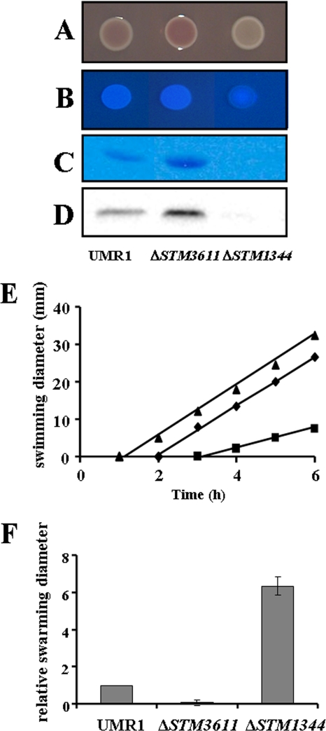
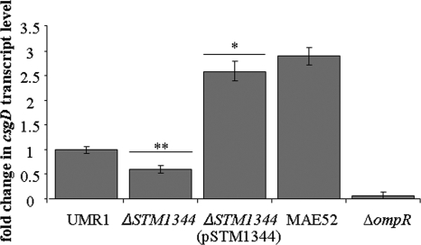
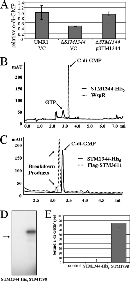
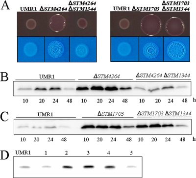
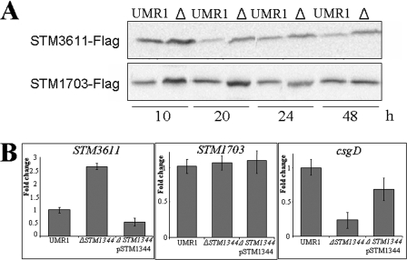
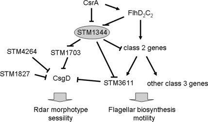
The bacterial second messenger cyclic di-GMP (c-di-GMP) regulates the transition between sessility and motility. In Salmonella enterica serovar Typhimurium, the expression of CsgD, the regulator of multicellular rdar morphotype behavior, is a major target of c-di-GMP signaling. CsgD expression is positively regulated by at least two diguanylate cyclases, GGDEF domain proteins, and negatively regulated by at least four phosphodiesterases, EAL domain proteins. Here, we show that in contrast to EAL domain proteins acting as phosphodiesterases, the EAL-like protein STM1344 regulated CsgD expression positively and motility negatively. STM1344, however, did not have a role in c-di-GMP turnover and also did not bind the nucleotide. STM1344 acted upstream of the phosphodiesterases STM1703 and STM3611, previously identified to participate in CsgD downregulation, where it repressed their expression. Consequently, although STM1344 has not retained a direct role in c-di-GMP metabolism, it still participates in the regulation of c-di-GMP turnover and has a role in the transition between sessility and motility.


Figures

FIG. 1.
FIG. 1.
Comparison of the amino acid sequence of the EAL-like protein STM1344 to that of the most similar EAL domain protein, the phosphodiesterase STM3611 in S. Typhimurium UMR1. Both proteins have only an EAL domain, with the name-giving EAL motif changed to EII in STM1344 and ELL in STM3611. Amino acid alignment was performed using the FFAS03 pairwise alignment server (14) and manual adjustment. Amino acids conserved in STM1344 and STM3611 are shown with a black background. The consensus motifs for functional phosphodiesterases are displayed below the alignment and shown with a gray background. The asterisks indicate alanine replacement of amino acids in STM1344, whereby only F168 was shown to have a functional role.
FIG. 2.
FIG. 2.
Phenotypes of the STM1344 knockout mutant in comparison with those of wild-type S. Typhimurium UMR1 and the c-di-GMP-specific phosphodiesterase STM3611 knockout mutant. (A to D) The expression of the rdar morphotype (A), calcofluor white binding capability (indicative of cellulose expression) (B), extracellular matrix component curli fimbriae (C), and the master regulator of rdar morphotype expression, CsgD (D), in the ΔSTM1344 mutant was downregulated in comparison to that in wild-type S. Typhimurium UMR1. Cells were grown on plates of LB agar without salt supplemented with Congo red (A) or calcofluor white (B) at 28°C for 20 h. The expression of CsgD was evaluated by Western blot analysis. Curli fimbriae were visualized by Coomassie brilliant blue staining of protein gels. (E and F) The swimming (E) and swarming (F) motilities of the ΔSTM1344 mutant were upregulated in comparison to those of wild-type S. Typhimurium UMR1. Diameters of the zones covered by swimming and swarming bacteria were measured. The regulation of all phenotypes of the ΔSTM1344 mutant was opposite that of the phenotypes of the STM3611 knockout mutant. Plates were incubated at 37°C for up to 6 h. In panel E, diamonds represent the wild-type strain UMR1, squares represent the STM3611 mutant, and triangles represent the STM1344 mutant.
FIG. 3.
FIG. 3.
Complementation of an S. Typhimurium UMR1 ΔSTM1344 mutant with wild-type and variant STM1344-His6 proteins. The complementation of ΔSTM1344 with a pBAD30 vector expressing STM1344-His6 (pSTM1344) leads to the upregulation of the rdar morphotype. (A) Calcofluor white binding and CsgD expression. (B) Complementation of ΔSTM1344 with STM1344-His6 variants. Only the F168A mutant of STM1344 did not complement CsgD expression. The N85A STM1344 variant produced a hypercomplementation phenotype. For Western blot analysis (A and B), cells were grown for 20 h on plates of LB agar without salt at 28°C. Pictures of plate-grown cells were taken after 48 h to make morphotype changes strongly visible. (C) Repression of swarming motility by the overexpression of STM1344-His6 and selected variants. While the N85A variant of STM1344 repressed swarming motility as did wild-type STM1344, the F164 variant of STM1344 had no effect. VC, pBAD30 vector control.
FIG. 4.
FIG. 4.
Western blot analysis of growth phase-dependent expression of CsgD in the ΔSTM1344 mutant of S. Typhimurium UMR1. As in wild-type S. Typhimurium UMR1, there is growth phase-dependent expression of CsgD in the ΔSTM1344 mutant. The expression of CsgD in the ΔSTM1344 mutant is reduced approximately 50% compared to that in the wild type. The reference was S. Typhimurium UMR1 or the ΔSTM1344 mutant grown for 16 h; the negative control was S. Typhimurium MAE46 (ΔompR) grown for 24 h.
FIG. 5.
FIG. 5.
Effect of STM1344 on csgD transcription in S. Typhimurium UMR1. The deletion of the STM1344 gene downregulated the csgD transcript level to approximately 60%, as determined by quantitative real-time RT-PCR. RNA was isolated from cells grown at 28°C for 20 h on LB agar plates without salt. Average values with standard deviations are displayed. **, P ≤ 0.01; *, P ≤ 0.05. The negative control was S. Typhimurium MAE46 ΔompR; the positive control was MAE52, showing csgD expression enhanced threefold compared to that in UMR1 (31).
FIG. 6.
FIG. 6.
Role of STM1344 in c-di-GMP metabolism in S. Typhimurium UMR1. (A) The total cellular c-di-GMP concentration is downregulated in the ΔSTM1344 mutant compared to those in wild-type S. Typhimurium UMR1 and the ΔSTM1344 mutant complemented with the STM1344-His6 sequence cloned into pBAD30. Cells were grown for 20 h on LB agar plates without salt at 28°C. Averages with standard deviations, which are based on two independent experiments with three technical replicates, are displayed. VC, pBAD30 vector control. (B to E) Purified STM1344 does not exhibit c-di-GMP-metabolizing and c-di-GMP binding activities. (B) Purified STM1344 does not exhibit diguanylate cyclase activity, in contrast to the positive control, constitutively active WspR* (18). Protein at 5 μM was incubated with 100 μM GTP for 1 h. The HPLC elution profiles of the reaction products are as follows: c-di-GMP elutes at 3.35 ml, whereas GTP elutes before 3 ml. The dashed-line curve represents the WspR* sample, whereas the solid-line curve represents the STM1344 sample. (C) Purified STM1344 does not exhibit c-di-GMP-specific phosphodiesterase activity, in contrast to the positive control, STM3611 (38). Protein at 1 μM was incubated with 100 μM c-di-GMP for 1 h at 37C. The HPLC elution profiles of the reaction products are as follows: c-di-GMP elutes at 3.35 ml; the first breakdown product, GpGp, elutes at 3.25 ml; and GMP elutes at 3.15 ml. The dashed-line curve represents the STM3611 sample, whereas the solid-line curve represents the STM1344 sample. mAU, milli-absorbance units. (D and E) Purified STM1344 does not show c-di-GMP binding, in contrast to the positive control, STM1798 (YcgR) (34), as determined by UV cross-linking (D) and equilibrium dialysis (E). The arrow in panel D indicates the position of STM1344-His6 UV cross-linking was performed with c-di-[32P]GMP. Protein at 10 μM was incubated with 100 μM c-di-GMP. For equilibrium dialysis, 200 pmol of c-di-GMP or other tested nucleotides was mixed with the purified proteins (200 pmol) in a dialysis cell surrounded by a membrane with an 8-kDa molecular mass cutoff. Equilibrium was reached after 16 h at room temperature with slow shaking. Average values with standard deviations are displayed.
FIG. 7.
FIG. 7.
Position of STM1344 in the c-di-GMP signaling network regulating CsgD expression in S. Typhimurium UMR1. (A) Expression of the rdar morphotype (top) and the calcofluor white binding phenotype (bottom) in wild-type S. Typhimurium UMR1, the corresponding mutants with knockouts of the STM4264 and STM1703 phosphodiesterase genes, and the respective double mutants with STM1344 deleted. While the knockout of STM1344 downregulated rdar morphotype expression in the STM4264 mutant background, only slightly altered rdar morphotype expression in the STM1703 background was observed. Cells were grown for 20 h at 28°C on plates of LB agar without salt, supplemented with Congo red or calcofluor white. (B to D) Effects of STM1344 deletion on CsgD expression in the STM4264 (B), STM1703 (C), STM3611 (D), and STM1827 (D) mutant backgrounds. CsgD was detected by Western blot analysis. Cells were grown on LB agar plates without salt at 28°C. In the analysis presented in panel D, cells were grown for 20 h. Lanes: 1, ΔSTM1344 mutant; 2, ΔSTM3611 mutant; 3, ΔSTM1344 ΔSTM3611 double mutant; 4, ΔSTM1827 mutant; 5, ΔSTM1344 ΔSTM1827 double mutant.
FIG. 8.
FIG. 8.
Effect of STM1344 knockout in S. Typhimurium UMR1 on the expression of the phosphodiesterases STM3611 and STM1703. (A) STM1344 downregulates the expression of STM3611 and STM1703 throughout the growth phase, as upregulated expression of STM3611 and STM1703 in the ΔSTM1344 mutant is observed. The STM3611 and STM1703 genes on the chromosome were linked with a FLAG tag sequence, and the proteins were detected with a FLAG antibody by Western blot analysis. Cells were grown on LB agar plates without salt at 28°C for the indicated times. Δ indicates the STM1344 knockout mutant of S. Typhimurium UMR1. (B) STM1344 affects the STM3611 gene transcript level but does not affect the STM1703 gene transcript level as determined by quantitative real-time RT-PCR. The csgD transcript was monitored as a positive control. RNA was isolated from cells grown for 16 h on LB agar plates without salt at 28°C. Average values with standard deviations are displayed.
FIG. 9.
FIG. 9.
Model illustrating the position of STM1344 in the c-di-GMP signaling network regulating the transition between sessility and motility. STM1344 positively affects the expression of the major regulator of the multicellular rdar morphotype behavior, CsgD, through the downregulation of the expression of the phosphodiesterases STM1703 and STM3611 (37). However, the regulation of STM3611 by STM1344 is probably indirect, as STM1344 represses not only STM3611, but also transcription from class 2 and class 3 promoters of the flagellar regulon (45). On the other hand, the expression of CsrA and the FlhD2C2 activator complex (expressed by the class 1 promoters) at the top of the flagellar regulon hierarchy downregulates STM1344 expression (; Jonas et al., unpublished). In addition, CsrA positively affects FlhD2C2 (44). STM4264 and STM1827 are two other phosphodiesterases that downregulate the expression of CsgD independently of STM1344 (37).

Similar articles

Cited by

References

    1. Ausubel, F. M., R. Brent, R. E. Kingston, D. D. Moore, J. G. Seidman, J. A. Smith, and K. Struhl (ed.). 1994. Current protocols in molecular biology. John Wiley & Sons, New York, NY.
    1. Brombacher, E., A. Baratto, C. Dorel, and P. Landini. 2006. Gene expression regulation by the curli activator CsgD protein: modulation of cellulose biosynthesis and control of negative determinants for microbial adhesion. J. Bacteriol. 1882027-2037. - PMC - PubMed
    1. Christen, M., B. Christen, M. Folcher, A. Schauerte, and U. Jenal. 2005. Identification and characterization of a cyclic di-GMP-specific phosphodiesterase and its allosteric control by GTP. J. Biol. Chem. 28030829-30837. - PubMed
    1. Cotter, P. A., and S. Stibitz. 2007. c-di-GMP-mediated regulation of virulence and biofilm formation. Curr. Opin. Microbiol. 1017-23. - PubMed
    1. Datsenko, K. A., and B. L. Wanner. 2000. One-step inactivation of chromosomal genes in Escherichia coli K-12 using PCR products. Proc. Natl. Acad. Sci. USA 976640-6645. - PMC - PubMed

Publication types

MeSH terms

LinkOut - more resources