. 2009 Jul;297(1):L52-63.
doi: 10.1152/ajplung.90406.2008. Epub 2009 Apr 17.

Novel critical role of Toll-like receptor 4 in lung ischemia-reperfusion injury and edema

Affiliations

Novel critical role of Toll-like receptor 4 in lung ischemia-reperfusion injury and edema

Giorgio Zanotti et al. Am J Physiol Lung Cell Mol Physiol. 2009 Jul.

Abstract

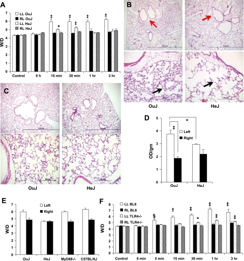
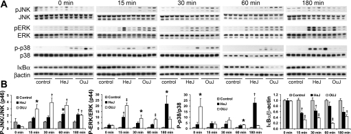
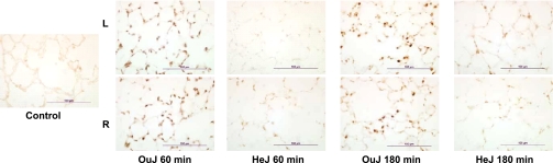
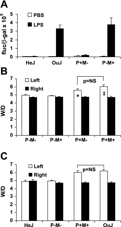
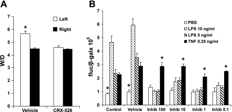
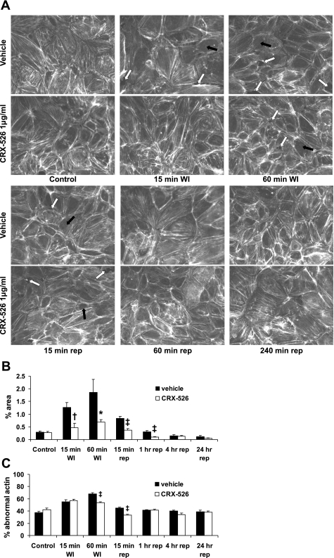
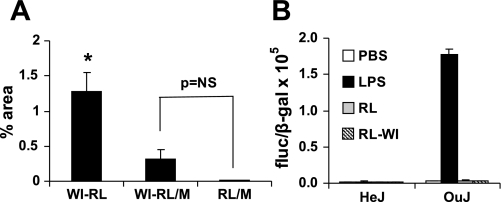
Toll-like receptors (TLRs) of the innate immune system contribute to noninfectious inflammatory processes. We employed a murine model of hilar clamping (1 h) with reperfusion times between 15 min and 3 h in TLR4-sufficient (C3H/OuJ) and TLR4-deficient (C3H/HeJ) anesthetized mice with additional studies in chimeric and myeloid differentiation factor 88 (MyD88)- and TLR4-deficient mice to determine the role of TLR4 in lung ischemia-reperfusion injury. Human pulmonary microvascular endothelial monolayers were subjected to simulated warm ischemia and reperfusion with and without CRX-526, a competitive TLR4 inhibitor. Functional TLR4 solely on pulmonary parenchymal cells, not bone marrow-derived cells, mediates early lung edema following ischemia-reperfusion independent of MyD88. Activation of MAPKs and NF-kappaB was significantly blunted and/or delayed in lungs of TLR4-deficient mice as a consequence of ischemia-reperfusion injury, but edema development appeared to be independent of activation of these signaling pathways. Pretreatment with a competitive TLR4 inhibitor prevented edema in vivo and reduced actin cytoskeletal rearrangement and gap formation in pulmonary microvascular endothelial monolayers subjected to simulated warm ischemia and reperfusion. In addition to its well-accepted role to alter gene transcription, functioning TLR4 on pulmonary parenchymal cells plays a key role in very early and profound pulmonary edema in murine lung ischemia-reperfusion injury. This may be due to a novel mechanism: regulation of endothelial cell cytoskeleton affecting microvascular endothelial cell permeability.


Figures

Fig. 1.
Fig. 1.
Toll-like receptor 4 (TLR4) is a key mediator of pulmonary edema [↑ wet-to-dry weight ratio (W/D)] due to ischemia-reperfusion injury (IRI). A: ischemia alone (0-h reperfusion) caused no increase in W/D. Following reperfusion, less edema develops in TLR4-deficient mice (HeJ), and is resolved by 1 h, but edema persists for 3 h in TLR4-sufficient mice (OuJ). LL, left lung; RL, right lung. *P < 0.05, ‡P < 0.001 compared with respective controls. P < 0.001 OuJ left lung compared with other lungs at 15-min, 30-min, 1-h, and 3-h reperfusion. n = 6/Group. ANOVA with Tukey post hoc. B: inflation-fixed (25 cmH2O) left lungs retrieved after 1-h hilar clamp and 3 h of reperfusion show increased interstitial edema in peribronchial and perivascular spaces in TLR4-sufficient mice (OuJ) compared with TLR4-deficient mice (HeJ) (red arrows; top; ×40) and thicker alveolar walls (black arrows; bottom; ×200). Representative of 4 specimens. C: despite significant difference in W/D following 60-min reperfusion, there is no interstitial peribronchial/perivascular edema in TLR4-sufficient (OuJ) and TLR4-deficient (HeJ) mice (top; ×40) and no alveolar wall thickening (bottom; ×200). Representative of 4 specimens. These inflation-fixed sections look identical to control specimens (data not shown). D: Evans blue dye accumulation in left lungs retrieved after 1 h of hilar clamping and 1 h of reperfusion supports the assumption that alveolar flooding is the reason for ↑W/D in TLR4-sufficient mice (OuJ) compared with TLR4-deficient mice (HeJ). *P < 0.05, unpaired t-test; ‡P < 0.05, paired t-test. OD, optical density. E: left lungs retrieved after 1 h of hilar clamping and 1 h of reperfusion from myeloid differentiation factor 88-deficient (MyD88−/−) mice (n = 5) develop the same increase in W/D as TLR4-sufficient (OuJ) mice (n = 6) and C57BL/6J mice (BL6; n = 6), their background strain. F: TLR4−/− mice develop significantly less edema than the background strain C57BL/6J mice after 1-h left hilar clamp and reperfusion for 5, 15, 30, 60, or 180 min (n = 3 at 5, 15, 30, 180 min, n = 6 otherwise). *P < 0.05, †P < 0.01, ‡P < 0.001, ANOVA with Tukey post hoc compared with respective controls; §P < 0.05 compared with other lungs at 5 min. For all other reperfusion times, P < 0.001, BL6 left lung compared with all others, ANOVA with Tukey post hoc. Although TLR4−/− left lungs gain some weight with time, they are not different compared with right lungs of either strain at any time point except at 1-h reperfusion (P < 0.05). This small weight gain with time may be due to strain differences in susceptibility to lung injury (see text). Bars in B and C: top, 2.0 mm; bottom, 200 μm.
Fig. 2.
Fig. 2.
Activation of MAPKs and NF-κB due to IRI differs between strains. A: protein extracted from left lungs rendered ischemic × 1 h and then reperfused for 0, 15, 30, 60, or 180 min. Activation of JNK, ERK, p38, and NF-κB is apparent early in TLR4-sufficient mice (OuJ), whereas TLR4-deficient mice (HeJ) have minimal or no degradation of JNK, reduced activation of ERK until 180 min, and very delayed activation of p38. IκBα degradation occurs earlier in OuJ mice and later (180 min) in HeJ mice. n = 4 Each of HeJ and OuJ strains; controls, 2 HeJ, 2 OuJ mice euthanized and lungs retrieved expeditiously. Protein from controls were placed on each gel for each time point to allow comparison with normal lung for each time point. B: quantification of intensity by laser scanning. Phospho-/total MAPKs and IκBα/β-actin were normalized by dividing each ratio by the mean ratio for controls. This makes each control = 1.0 with variability among the different control samples represented by error bars. Values are means ± SE. p46 and p54 JNK and p44 and p42 ERK have similar patterns and P values. *P < 0.05, †P < 0.01, ‡P < 0.001 compared with controls by ANOVA with Tukey honestly significant difference (HSD) for multiple comparisons. p, Phosphorylated.
Fig. 3.
Fig. 3.
Immunostaining for p65 component of NF-κB. Minimal nuclear localization in control (freshly euthanized) with marked nuclear staining (3+) in 60-min reperfused samples in TLR4-sufficient (OuJ) mice compared with TLR4-deficient (HeJ) strain where staining was graded 1–2+. Immunostaining intensity complemented IκBα degradation (Fig. 2) except that IκBα levels appear equivalent in HeJ and OuJ strains at 180-min reperfusion despite more p65 staining in OuJ animals at 180-min reperfusion. We interpret this to mean some recovery of IκBα protein in OuJ mice 180 min postreperfusion. Curiously, p65 staining was the same for right (R) and left (L) lungs in both strains, implying equivalent NF-κB activation in both lungs following reperfusion of the left ischemic lung. Bars = 100 μm.
Fig. 4.
Fig. 4.
Functioning TLR4 on lung parenchymal cells is necessary for development of edema due to IRI. P, parenchymal cells; M, marrow-derived cells; +, intact TLR4 (OuJ); −, nonfunctional TLR4 (HeJ). A: alveolar macrophages (AMs) retrieved by bronchoalveolar lavage (BAL) were infected with Ad.NFκBLuc and Ad.CMV-LacZ and then incubated with 1 μg/ml PBS or LPS. Firefly luciferase/β-galactosidase (fluc/β-gal) activity shows complete replacement of recipient marrow from either HeJ strain (P+M−) or OuJ strain (P−M+). AMs retrieved from nonirradiated HeJ and OuJ mice served as controls. LPS stimulation resulted in ∼60-fold increase in luciferase activity in both native OuJ AMs and in AMs retrieved from chimeric strain P−M+. n = 4 Experiments/group; P < 0.0001. AMs retrieved from irradiated mice reconstituted with same strain marrow behaved in the same manner (data not shown). B: functioning TLR4 on parenchymal cells (P) is necessary for development of edema after 3-h IRI. P+M− chimeras developed significant increase in W/D. However, even if AMs had functioning TLR4 (P−M+), W/D was not elevated. Thus functioning TLR4 on AMs is not sufficient for development of edema but may amplify edema in mice with functioning TLR4 on lung parenchymal cells; W/D was slightly higher in P+M+ animals compared with P+M−, but the difference was not significant (NS). n = 5/Group; *P < 0.05, †P < 0.01 compared with W/D of P− left lungs (ANOVA with Tukey HSD). C: chimeras with restored bone marrow (P−M−, P+M+) had the same W/D as the intact strains (OuJ, HeJ) demonstrating that lethal radiation and bone marrow transplant (BMT) had no effect on development of edema due to IRI.
Fig. 5.
Fig. 5.
A competitive inhibitor (Inhib) of TLR4 (CRX-526) prevents lung edema due to IRI and TLR4-mediated activation of NF-κB. A: W/D of left lungs of OuJ mice pretreated with vehicle or 10 μg CRX-526 in 200 μl of normal saline administered over 30 min, 30 min before left hilar clamping for 1 h and 1-h reperfusion. Mice pretreated with CRX-526 had the same W/D as HeJ mice 1 h postreperfusion in Fig. 1A. n = 5/Group; *P = 0.0014 compared with right lung of same animal by paired t-test; P = 0.0023 compared with left lung of mice pretreated with CRX-526 by unpaired t-test. B: concentrations of CRX-526 from 100 μg/ml to 0.1 μg/ml successfully inhibited NF-κB activation (based on luciferase/β-gal activity) following stimulation of human pulmonary microvascular endothelial cells (HMVECs) in 96-well plates with LPS (10 or 5 ng/ml), but CRX-526 had no impact on TNF stimulation of NF-κB activation (black bars). n = 4/Group; *P < 0.05 compared with other values at same time point by ANOVA.
Fig. 6.
Fig. 6.
Simulated warm ischemia (WI) without hypoxia causes actin cytoskeletal rearrangement and formation of gaps in the human pulmonary microvascular endothelial monolayer. A: HMVECs grown to confluence on P30 dishes with integral coverslips were incubated with 1 μg/ml CRX-526 or vehicle and ventilated with 95% O2-5% CO2. Media was replaced with warm (37°C) Ringer lactate (RL) and ventilated with 100% O2 to simulate warm ischemia. One hour later, RL was replaced with warm cell culture media, and chambers were ventilated with 95% room air-5% CO2 to simulate reperfusion. During simulated warm ischemia, actin stress fibers disappeared or became more peripheral in the cells (black arrows), associated with formation of gaps in the endothelial monolayer (white arrows). Four hours after simulated reperfusion (240 min rep) and 24 h after simulated reperfusion (data not shown), monolayers were confluent, and actin cytoskeleton pattern was similar to controls. Gaps in the monolayer and actin cytoskeletal rearrangement were reduced by CRX-526. Experiments were performed in triplicate. B: %area of gaps in the monolayer (quantified by MetaMorph software) was reduced by CRX-526. Three separate fields from each of 3 P30 dishes were analyzed (n = 9 photos/time point). *P < 0.05, †P < 0.01, ‡P < 0.001, unpaired t-test. C: distributions of actin cytoskeletal rearrangement at different time points in simulated warm IRI. Because of considerable variability in the actin cytoskeleton of HMVECs, cells were labeled as normal or abnormal actin distribution in images (n = 9 photos/time point) without attempting to grade the severity of the abnormality. The assessment was made by a masked observer unaware of the group identity or the time of the sample, and then ratios of populations were calculated. Approximately 40% of cells have some degree of peripheral orientation of the actin cytoskeleton in fresh control dishes. In cells subjected to simulated warm ischemia, peripheral orientation of actin was significantly reduced in the presence of TLR4 inhibitor CRX-526 following 60 min of WI and after 15-min simulated reperfusion compared with monolayers exposed to vehicle. ‡P < 0.001, unpaired t-test.
Fig. 7.
Fig. 7.
TLR4-mediated alteration of HMVEC cytoskeleton due to IRI is ligand-independent. A: quantification of gap area by MetaMorph software. WI-RL, media replaced by RL for 60 min; WI-RL/M, RL from this ischemia experiment diluted with equal volume of fresh media; RL/M, fresh RL mixed with equal volume of fresh media (glucose concentration is halved). HMVECs exposed for 60 min to WI-RL or RL/M do not develop significant gaps compared with HMVECs when media is replaced with RL (WI-RL). *P < 0.01. B: RL removed from HMVECs following 1 h of simulated ischemia (RL-WI) and placed on transfected AMs retrieved from HeJ and OuJ mice (n = 4 each). There is no activation of NF-κB by this RL-WI or fresh RL.

Similar articles

Cited by

References

    1. Casiraghi M, Abano J, Burridge K, Randell S, Egan T. Toll-like receptor-4 (TLR4) inhibition reduces actin cytoskeletal re-arrangement and gap formation in cultured human pulmonary microvascular endothelial cells (HMVECs) subjected to simulated warm ischemia-reperfusion injury (IRI) (Abstract). J Heart Lung Transplant 27: S214–S215, 2008.
    1. Cho HY, Kleeberger SR. Genetic mechanisms of susceptibility to oxidative lung injury in mice. Free Radic Biol Med 42: 433–445, 2007. - PubMed
    1. Chow JC, Young DW, Golenbock DT, Christ WJ, Gusovsky F. Toll-like receptor-4 mediates lipopolysaccharide-induced signal transduction. J Biol Chem 274: 10689–10692, 1999. - PubMed
    1. de Groot H, Rauen U. Ischemia-reperfusion injury: processes in pathogenetic networks: a review. Transplant Proc 39: 481–484, 2007. - PubMed
    1. de Perrot M, Liu M, Waddell TK, Keshavjee S. Ischemia-reperfusion-induced lung injury. Am J Respir Crit Care Med 167: 490–511, 2003. - PubMed

Publication types

MeSH terms