. 2009 Jun;15(6):1121-33.
doi: 10.1261/rna.1195709. Epub 2009 Apr 17.

Vg1RBP phosphorylation by Erk2 MAP kinase correlates with the cortical release of Vg1 mRNA during meiotic maturation of Xenopus oocytes

Affiliations

Vg1RBP phosphorylation by Erk2 MAP kinase correlates with the cortical release of Vg1 mRNA during meiotic maturation of Xenopus oocytes

Anna Git et al. RNA. 2009 Jun.

Abstract

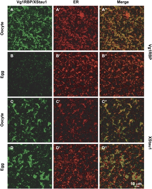
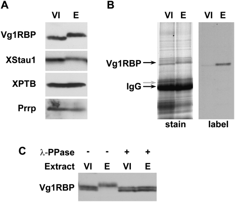
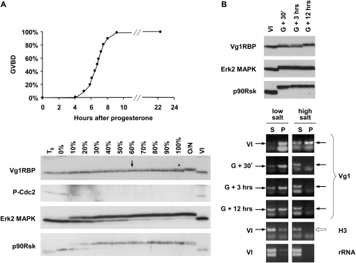
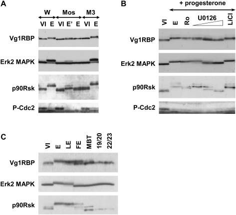
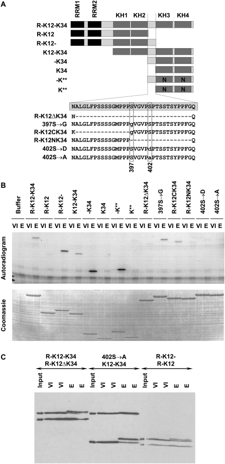
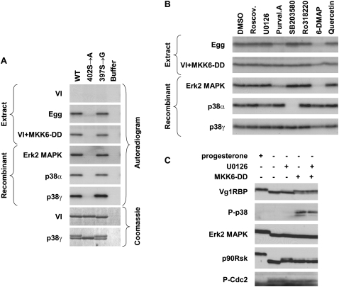
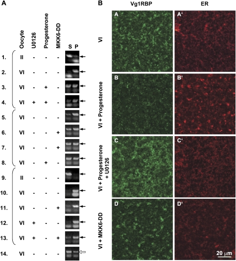
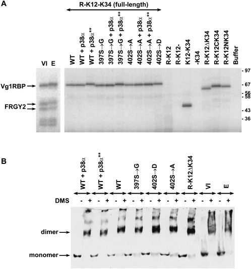
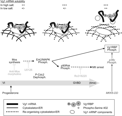
Xenopus Vg1RBP is a member of the highly conserved IMP family of four KH-domain RNA binding proteins, with roles in RNA localization, translational control, RNA stability, and cell motility. Vg1RBP has been implicated in localizing Vg1 mRNAs to the vegetal cortex during oogenesis, in a process mediated by microtubules and microfilaments, and in migration of neural crest cells in embryos. Using c-mos morpholino, kinase inhibitors, and constitutely active recombinant kinases we show that Vg1RBP undergoes regulated phosphorylation by Erk2 MAPK during meiotic maturation, on a single residue, S402, located between the KH2 and KH3 domains. Phosphorylation temporally correlates with the release of Vg1 mRNA from its tight cortical association, assayed in lysates in physiological salt buffers, but does not affect RNA binding, nor self-association of Vg1RBP. U0126, a MAP kinase inhibitor, prevents Vg1RBP cortical release and Vg1 mRNA solubilization in meiotically maturing eggs, while injection of MKK6-DD, a constitutively activated MAP kinase kinase, promotes the release of both Vg1RBP and Vg1 mRNA from insoluble cortical structures. We propose that Erk2 MAP kinase phosphorylation of Vg1RBP regulates the protein:protein-mediated association of Vg1 mRNP with the cytoskeleton and/or ER. Since the MAP kinase site in Vg1RBP is conserved in several IMP homologs, this modification also has important implications for the regulation of IMP proteins in somatic cells.


Figures

FIGURE 1.
FIGURE 1.
Vg1RBP distribution in the vegetal subcortical region of oocytes and eggs. Confocal images of stage VI oocytes and progesterone-matured eggs stained with anti-Vg1RBP (A,B) and anti-XStau1 (C,D) antibodies. All samples were costained with anti-GRP94 antibodies to visualize the ER (A′–D′). (A″–D″) are the corresponding overlays of Vg1RBP/XStau1 (green) and GRP94 (red). The confocal sections were taken about 5 μm below the vegetal surface in a layer containing distinctive patches containing Vg1RBP and XStau1. Scale bar 10 μm.
FIGURE 2.
FIGURE 2.
Vg1RBP is phosphorylated during meiotic maturation. (A) Vg1RBP undergoes an apparent shift in mobility during meiotic maturation. Protein samples from stage VI oocytes (VI) or progesterone-matured eggs (E) were analyzed by Western blotting using antisera directed against the proteins indicated on the left. (B) Vg1RBP is phosphorylated during meiotic maturation. Stage VI oocytes were incubated overnight in the presence (E) or absence (VI) of progesterone in a medium containing 33P orthophosphate. Vg1RBP was immunoprecipitated using anti-Vg1RBP antiserum and visualized either by autoradiography (right) or Coomassie Blue staining (left). Note that the overall immunoprecipitation efficiency is similar in both samples. The migration of Vg1RBP and of the IgG heavy chain is indicated. (C) The modification of Vg1RBP is phosphatase-sensitive. Protein samples from stage VI oocytes (VI) or progesterone-matured eggs (E) were treated with λ-phosphatase (λ-ppase) and compared to untreated samples using Western blot analysis. Note that under the PAGE conditions employed for this experiment, unmodified Vg1RBP migrates as doublet, reflecting the two allelic variants, A and D.
FIGURE 3.
FIGURE 3.
Vg1RBP phosphorylation is a late event in meiotic maturation and correlates with release of Vg1 mRNA into the soluble fraction. (A) Full phosphorylation of Vg1RBP is a late event in meiotic maturation. Random groups of 10 cells out of ∼150 stage VI oocytes treated with progesterone were taken at the time of progesterone addition (T0), when the first GVBD was observed (0%), when the indicated proportion of cells has undergone GVBD (10%–100%) and after an overnight incubation with progesterone (O/N). Control oocytes were incubated for the duration of the experiment in the absence of progesterone (VI). Oocytes were then subjected to Western blot analysis using antisera directed against the proteins indicated on the left. Asterisk: ∼40% phosphorylation; arrow: earliest visible phospho-Vg1RBP. (B) Phosphorylation of Vg1RBP coincides with solubilization of Vg1 mRNA in low-salt conditions. Groups of synchronized stage VI oocytes undergoing maturation were harvested 30 min (G + 30′), 3 h (G + 3 h), or 12 h (G + 12 h) after synchronization. Control oocytes were incubated for the duration of the experiment in the absence of progesterone (VI). Oocytes were then either subjected to Western blot analysis using antisera directed against the indicated proteins (top panel) or fractionated into soluble (S) and insoluble (P) fractions in different buffers (see Experimental Procedures for more details). Equivalent proportions of RNA extracted from these fractions was then assayed for the presence of Vg1 and histone H3 mRNAs using nonsaturated RT-PCR. Arrows indicate the diagnostic PCR product. The yield and integrity of extracted RNA was examined by electrophoresis in the presence of ethidium bromide (rRNA).
FIGURE 4.
FIGURE 4.
Vg1RBP phosphorylation is dependent on the MAPK pathway. (A) Inhibition of Mos synthesis prevents Vg1RBP phosphorylation. Stage VI oocytes were injected with 50 nL of water (W), 0.5 mM anti-Mos morpholino antisense (Mos), or 0.5 mM control morpholino oligonucleotide (M3), and incubated overnight in the presence (E, E′; duplicate samples) or absence (VI) of progesterone. Cells were then subjected to Western blot analysis using antisera directed against the proteins indicated on the left. (B) Inhibition of Mek1, but not p90Rsk or GSK3β, prevents phosphorylation of Vg1RBP. Stage VI oocytes were preincubated with 5 μM Ro318220, 10, 20, or 50 μM U0126, 10 mM LiCl, or DMSO at comparable concentrations for 4 h and then left overnight in the presence or absence (VI) of progesterone. Cells were then subjected to Western blot analysis using antisera directed against the proteins indicated on the left. (C) Vg1RBP phosphorylation in embryogenesis correlates with MAPK activity. Protein equivalents of one-stage VI oocyte (VI), progesterone-matured egg (E), laid egg (LE), fertilized egg (FE), MBT embryo, and stage 19/20 or 22/23 neurulas were subjected to Western blot analysis using antisera against the proteins indicated on the left.
FIGURE 5.
FIGURE 5.
Vg1RBP is phosphorylated on Serine 402. (A) Schematic representation of recombinant Vg1RBP variants and their nomenclature. Black: RRMs, dark gray: KH domains, light gray: unstructured sequences, N: point mutations in KH domains which abrogate RNA binding. The wild-type sequence of the inter-KH linker (spanning residues 381–414) is boxed; internal deletions are showed as hyphens, point mutations are in lowercase, the position of serines 397 and 402 is indicated below. The drawing is not to scale. (B) Serine 402 is phosphorylated in vitro; 15 pmol of each of the indicated Vg1RBP variants was used as a substrate in an in vitro kinase assay using extracts derived from either stage VI oocytes (VI) or progesterone-matured eggs (E). Samples were visualized using autoradiography (top panel) and Coomassie Blue staining (bottom panel). (C) Serine 402 is phosphorylated in vivo. Pairs of equimolar recombinant Vg1RBP variants were injected into stage VI oocytes, which were then incubated overnight in the presence (E) or absence (VI) of progesterone. Protein extracts from duplicate samples were subjected to Western blot analysis using anti-His antibody alongside samples of the injected mixtures (input). Injected proteins are indicated with the larger protein above the smaller protein. Note that due to differential stability of the injected proteins, samples from R-K12/R-K12- injected cells represent two rather than one cell equivalents.
FIGURE 6.
FIGURE 6.
Vg1RBP is phosphorylated by MAPK. (A) Serine 402 can be phosphorylated by both Erk2 and p38 MAP kinases; 15 pmol of recombinant full-length wild-type (WT), S402 → A or S397 → G Vg1RBP variants or buffer alone were used as substrates for in vitro kinase assays with 10 ng recombinant in vitro-activated Erk2, p38α, or p38γ or extracts made from stage VI oocytes (VI), progesterone-matured eggs (E), and stage VI oocytes injected with MKK6-DD (VI + MKK6-DD). Samples were visualized using autoradiography and Coomassie Blue staining. (B) The phosphorylation of Vg1RBP by egg extract is sensitive to Erk2 MAPK but not p38/MAPK inhibitors; 15 pmol wild-type recombinant Vg1RBP was used as a substrate for in vitro kinase assays using kinases and extracts as above in the presence of the following inhibitors: 10 μM Roscovitine, 20 μM U0126, 10 μM Purvalanol A, 10 μM SB203580, 1 μM Ro328220, 1 mM 6-DMAP, or 20 μM Quercetin. A similar volume of DMSO was used as a control. (C) In vivo activation of p38 MAPK leads to Vg1RBP phosphorylation in the absence of cell cycle progression. Stage VI oocytes were preincubated for 4 h with 50 μM U0126 or equivalent concentrations of DMSO, microinjected with 50 nL of 1.5 mg/mL MKK6-DD and incubated overnight alongside with progesterone-treated oocytes. Cells were then subjected to Western blot analysis using antisera directed against the proteins indicated on the left.
FIGURE 7.
FIGURE 7.
Vg1RBP phosphorylation correlates with its cortical detachment and solubilization of Vg1 mRNA. (A) The solubility of Vg1 mRNA in low salt buffer correlates with the phosphorylation of Vg1RBP. Stage VI oocytes were preincubated with 50 μM U0126 where indicated and then either injected with 50 nL 1.5 mg/mL MKK6-DD or exposed to progesterone. Control stage II and VI oocytes were incubated for the duration of the experiment without any treatment. After a 16-h incubation, oocytes were fractionated into soluble (S) and insoluble (P) fractions in low salt buffer. Equivalent proportions of RNA extracted from these fractions was then assayed for the presence of Vg1 (samples 1–13) or histone H3 (sample 14) mRNAs using nonsaturated RT-PCR. Arrows indicate the diagnostic PCR product. Brackets group samples that originated from the same experiment. (B) Vg1RBP phosphorylation correlates with its cortical release. Confocal images of stage VI oocytes (VI), oocytes matured by addition of progesterone in the absence or presence of 50 μM U0126 and stage VI oocytes injected with MKK6-DD (VI + MKK6-DD), stained with anti-Vg1RBP (A–D) and anti-GRP94 antibodies (visualizing the ER; A′–D′). Confocal images from equivalent subcortical layers near the vegetal pole are shown, except for the MKK6-injected oocytes where the slightly deeper layer to which the ER-Vg1RBP patches have sunk is shown. Scale bar: 20 μm.
FIGURE 8.
FIGURE 8.
Vg1RBP phosphorylation does not affect its RNA binding or self-association. (A) Phosphorylation of Vg1RBP does not affect RNA binding. S10 extracts from stage VI oocytes (VI) or progesterone-matured eggs (E) and 0.75 pmol recombinant Vg1RBP variants were tested in a UV cross-linking assay using VLE RNA probe uniformly labeled with [α-32P]-UTP. Where indicated, proteins were prephosphorylated with recombinant in vitro activated p38α (+ p38α**). A parallel reaction with inactive p38α (+ p38α) was used as a control. The samples were resolved using SDS-PAGE and visualized by autoradiography. (B) Phosphorylation of Vg1RBP does not affect self association. S10 extracts from stage VI oocytes (VI) or progesterone-matured eggs (E) and 15 pmol recombinant Vg1RBP variants were cross-linked by addition of 1.5 mg/mL dimethyl suberimidate (DMS) in borate buffer (+) or borate buffer alone (−). Where indicated, proteins were prephosphorylated as above. Samples were resolved on phosphate SDS-PAGE gel and subjected to Western blot analysis using anti-Vg1RBP antiserum. Migration of monomer and dimer forms is indicated on the left.
FIGURE 9.
FIGURE 9.
A two-step model for the release of Vg1 mRNA from the cortex in meiotic maturation. Initiating triggers for the phosphorylation of Vg1RBP (boxed) are indicated at the bottom (application of progesterone or microinjection of MKK6-DD) with the physiological pathway arranged temporally (from left to right; not to scale). The components contributing to Vg1 mRNA's subcortical anchoring at the key stages are represented in the main illustration (see key in the figure). The solubility of Vg1 mRNA in high or low salt buffers is indicated above. The activity of several inhibitors used throughout this study is in light gray. Direct links are in solid arrows and indirect connections are in dashed arrows. Phosph., phosphorylation; R, RRMs; K, KH didomains.

Similar articles

Cited by

References

    1. Allison R., Czaplinski K., Git A., Adegbenro E., Stennard F., Houliston E., Standart N. Two distinct Staufen isoforms in Xenopus are vegetally localized during oogenesis. RNA. 2004;10:1751–1763. - PMC - PubMed
    1. Alonso G., Ambrosino C., Jones M., Nebreda A.R. Differential activation of p38 mitogen-activated protein kinase isoforms depending on signal strength. J. Biol. Chem. 2000;275:40641–40648. - PubMed
    1. Bain J., McLauchlan H., Elliott M., Cohen P. The specificities of protein kinase inhibitors: An update. Biochem. J. 2003;371:199–204. - PMC - PubMed
    1. Barbee S.A., Estes P.S., Cziko A.M., Hillebrand J., Luedeman R.A., Coller J.M., Johnson N., Howlett I.C., Geng C., Ueda R., et al. Staufen- and FMRP-containing neuronal RNPs are structurally and functionally related to somatic P bodies. Neuron. 2006;52:997–1009. - PMC - PubMed
    1. Becker B.E., Gard D.L. Visualization of the cytoskeleton in Xenopus oocytes and eggs by confocal immunofluorescence microscopy. Methods Mol. Biol. 2006;322:69–86. - PubMed

Publication types

LinkOut - more resources