. 2009 Apr;21(4):1252-72.
doi: 10.1105/tpc.109.065565. Epub 2009 Apr 17.

Misexpression of FATTY ACID ELONGATION1 in the Arabidopsis epidermis induces cell death and suggests a critical role for phospholipase A2 in this process

Affiliations

Misexpression of FATTY ACID ELONGATION1 in the Arabidopsis epidermis induces cell death and suggests a critical role for phospholipase A2 in this process

José J Reina-Pinto et al. Plant Cell. 2009 Apr.

Abstract

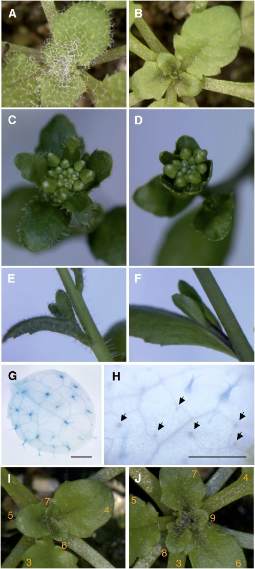
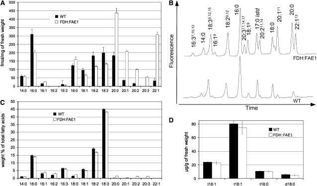
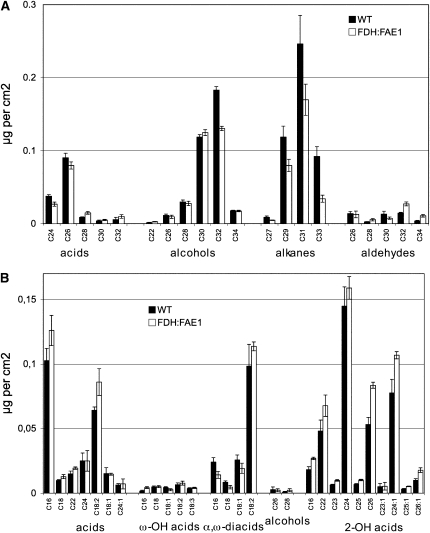
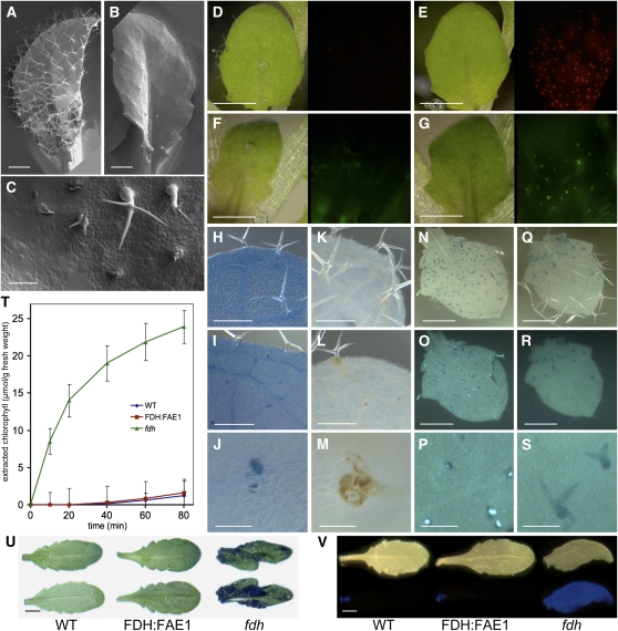
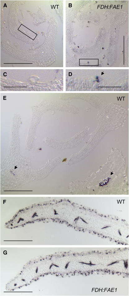
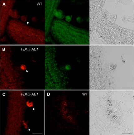
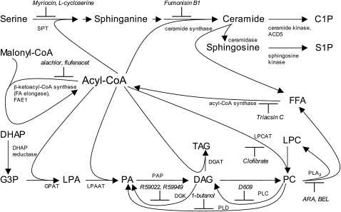
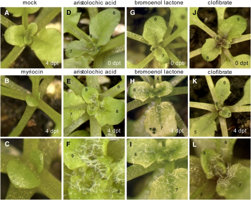
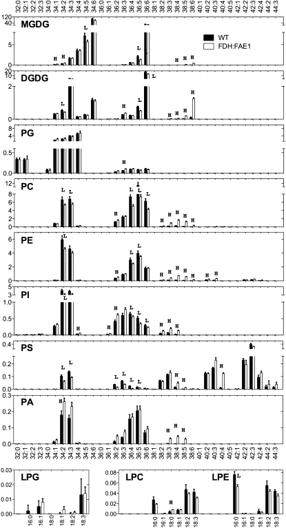
Very-long-chain fatty acids (VLCFAs) are important functional components of various lipid classes, including cuticular lipids in the higher plant epidermis and lipid-derived second messengers. Here, we report the characterization of transgenic Arabidopsis thaliana plants that epidermally express FATTY ACID ELONGATION1 (FAE1), the seed-specific beta-ketoacyl-CoA synthase (KCS) catalyzing the first rate-limiting step in VLCFA biosynthesis. Misexpression of FAE1 changes the VLCFAs in different classes of lipids but surprisingly does not complement the KCS fiddlehead mutant. FAE1 misexpression plants are similar to the wild type but display an essentially glabrous phenotype, owing to the selective death of trichome cells. This cell death is accompanied by membrane damage, generation of reactive oxygen species, and callose deposition. We found that nuclei of arrested trichome cells in FAE1 misexpression plants cell-autonomously accumulate high levels of DNA damage, including double-strand breaks characteristic of lipoapoptosis. A chemical genetic screen revealed that inhibitors of KCS and phospholipase A2 (PLA2), but not inhibitors of de novo ceramide biosynthesis, rescue trichome cells from death. These results support the functional role of acyl chain length of fatty acids and PLA2 as determinants for programmed cell death, likely involving the exchange of VLCFAs between phospholipids and the acyl-CoA pool.


Figures

Figure 1.
Figure 1.
Glabrous Phenotype of FDH:FAE1 Transgenic Plants. (A) to (F) Comparison of 3.5-week-old wild-type (A) and FDH:FAE1 (B) rosettes (grown under short-day conditions), wild-type (C) and FDH:FAE1 (D) inflorescences, and stems from 5-week-old wild-type (E) and FDH:FAE1 (F) plants carrying cauline leaves and axillary shoots. (G) and (H) Expression of the GL2:GUS reporter in developing leaves of 2-week-old wild-type (G) and FDH:FAE1 (H) seedlings. Note that most trichomes remained undeveloped and were not histochemically stained for GUS in the double transgenics (arrows). Bars = 500 μm. (I) and (J) Chemical rescue of trichome development with 0.005% alachlor. (I) and (J) show the same plant before and 1 week after exposure to alachlor, a potent inhibitor of KCSs. The inhibitor was applied to 3-week-old FDH:FAE1 seedlings by spraying, to inactivate FAE1 (I). Leaves were numbered from the base to the apex of the plant. Young leaves, which developed 1 week after spraying, exhibit trichomes (J).
Figure 2.
Figure 2.
Analysis of the Fatty Acid Composition of the Acyl-CoA and Total Lipid Pools and of LCBs in FDH:FAE1 and Wild-Type Plants. (A) HPLC analysis of VLCFAs in acyl-CoA esters (mean ± se; n = 5). Acyl-CoAs were extracted from young rosette leaves of 6-week-old plants (0.5 to 1.0 cm), derivatized to fluorescent acyl-etheno-CoAs, and analyzed by HPLC. (B) Representative HPLC profiles of acyl-CoAs in FDH:FAE1 and wild-type plants obtained as summarized in (A). (C) Gas chromatography (GC) analysis of total fatty acids isolated from the same tissues as in (A) (mean ± se; n = 5). Fatty acids were transesterified and analyzed as methyl esters (FAMEs). (D) LCBs were extracted and analyzed as dinitrophenyl derivatives by reverse-phase HPLC/MS (mean ± se; n = 4). In the figure, “d” denotes dihydroxy LCBs, and “t” denotes trihydroxy LCBs; the numbers designate the length and degree of desaturation of the acyl chain, respectively; t18:1(Z), 4-hydroxy-8-(cis)-sphingenine; t18:1(E), 4-hydroxy-8-(trans)-sphingenine; t18:0, 4-hydroxysphinganine; d18:0, dihydrosphinganine.
Figure 3.
Figure 3.
Composition Analysis of Epicuticular Waxes and Residual-Bound Lipids in Leaves. (A) Wax was extracted by rapid dipping in chloroform and BSTFA derivatized and analyzed by GC and GC-MS (mean ± se; n = 5). Leaves of 6-week-old plants were harvested for analysis in (A) and (B). (B) For cell wall–bound lipid analysis, leaves were first extensively defatted with chloroform-methanol. Bound lipids were analyzed using GC and GC-MS as described previously (Franke et al., 2005; Kurdyukov et al., 2006a) (mean ± se; n = 5).
Figure 4.
Figure 4.
Characterization of the Trichome Cell Death Phenotype in FDH:FAE1 Plants. (A) and (B) Scanning electron micrographs showing the adaxial epidermis of rosette leaves. Note the fully developed trichomes on the wild-type leaf (A) and the arrested trichomes characteristic of FDH:FAE1 plants (B). Bars = 500 μm. (C) Close-up view of FDH:FAE1 trichomes, including some that arrested or underwent collapse and degeneration at various developmental stages. Bar = 100 μm. (D) and (E) Micrographs of young leaves (left panels) stained with PI and viewed using the DsRed filter set (right panels). Note that PI labels trichomes in the FDH:FAE1 leaf (E) but not the wild type (D). Bars = 500 μm. (F) and (G) Micrographs of young leaves (left panels) stained with DCFH-DA and viewed using the GFP filter set (right panels). The DCFH-DA fluorescence reveals the presence of ROS in FDH:FAE1 trichomes (G) but not in wild-type trichomes (F). Bars = 500 μm. (H) to (J) Trypan blue staining detects cell death in differentiating TEs in wild-type (H) and FDH:FAE1 (I) leaves and trichome death in FDH:FAE1 ([I] and [J]). Note that some mature trichomes in (I) are viable and are not stained. (J) shows detail of a FDH-FAE1 trichome. Bars = 250 μm in (H) and (I) and 50 μm in (J). (K) to (M) Wild-type (K) and FDH:FAE1 ([L] and [M]) leaves stained with DAB to detect hydrogen peroxide. A brown reaction product indicates the presence of H2O2 in trichomes in FDH:FAE1 and in the vasculature of both wild-type and FDH:FAE1 leaves. (M) shows detail of a FDH-FAE1 trichome. Bars = 250 μm in (K) and (L) and 50 μm in (M). (N) to (S) Aniline blue fluorescence test to detect callose. Bright blue-white fluorescent spots indicate the presence of callose in FDH:FAE1 trichomes ([O] and [P]) but not in wild-type leaves ([R] and [S]). (N) and (Q) are visible-light images, and (O) and (R) are UV-light images. (P) and (S) show enlarged details from (O) and (R), respectively. That the callose depositions are primarily localized to branch tips could be seen also in Supplemental Figure 2 online. Bars = 500 μm in (N), (O), (Q), and (R) and 50 μm in (P) and (S). (T) Kinetics of chlorophyll leaching from leaves of the indicated genotypes into ethanol solution. Leaves were incubated in 70% ethanol for the times indicated. Note that FDH:FAE1 leaves behave like the wild type, whereas fdh shows abnormal cuticle permeability. (U) Staining of leaves of wild-type, FDH:FAE1, and fdh plants with TB to reveal cuticular defects. Adaxial (top panel) and abaxial (bottom panel) sides of leaves are shown. Note that only fdh tissues were positively stained. Bar = 5 mm. (V) Calcofluor white test for cuticle permeability. Leaves of wild-type, FDH:FAE1, and fdh plants (top panel) were stained with calcofluor white and examined under UV light (bottom panel). The permeable fdh leaf was used as positive control; wild-type and FDH:FAE1 leaves were not stained; however, FDH:FAE1 dead trichomes could be visualized following an a more extended TB staining (see Supplemental Figure 3 online). Bar = 5 mm.
Figure 5.
Figure 5.
Detection of DNA Fragmentation in Leaves by in Situ TUNEL-LID. Relatively thick sections (14 μm) were cut to enhance the visibility of trichomes. DNA strand breaks were detected by enzymatically labeling the free 3′-OH, followed by hybridization of a specific digoxigenin-labeled probe to the added tag. Hybridization signals were visualized as blue-purple intranuclear precipitates. Bars = 200 μm in (A), (B), and (E) to (G) and 50 μm in (C) and (D). (A) and (B) Cross sections of shoot apexes at a nonflowering stage, showing leaf primordia in the wild type (A) and FDH:FAE1 (B). Note specific TUNEL labeling in FDH:FAE1 trichomes (B). (C) and (D) High-resolution views of the areas boxed in (A) and (B), respectively. In (B), the labeling of the FDH:FAE1 trichome (arrow) indicates the presence of DNA strand breaks. (E) Differentiating TEs in older wild-type leaves (arrows) labeled under the same conditions as used in (A) and (B). Nuclear fragmentation is a normal part of TE formation, which involves PCD. (F) and (G) Longer staining periods (up to overnight) result in a notable increase in sensitivity and reveal DNA strand breaks in the epidermis and developing vasculature of both wild-type (F) and FDH:FAE1 (G) leaves.
Figure 6.
Figure 6.
Detection of Blunt-End and Single 3′ dN Overhang DNA Fragments by in Situ Ligation. Confocal fluorescence images of ATTO 590 labeling (red) and EvaGreen staining (green) and transmission images (gray) of cross sections were obtained from FDH:FAE1 ([B] and [C]) and wild-type plants ([A] and [D]) at a nonflowering stage. EvaGreen preferentially stained the nuclear DNA and, to a lesser extent, cytoplasm due to indiscriminate staining of RNA. Bars = 20 μm. (A) Wild-type leaf primordium. Trichome nuclei are not labeled by the ATTO 590 oligonucleotide probe (arrow). (B) FDH:FAE1 leaf primordium showing bright ATTO 590 labeling of fragmented chromatin (arrow), which has undergone condensation at the periphery of the trichome nucleus. Note the absence of ATTO 590 labeling in other epidermal cells in (A) and (B). (C) FDH:FAE1 leaf showing disorganized chromatin labeled with the ATTO 590 probe in two trichomes (arrows). (D) ATTO 590 fluorescence and transmission images of vascular bundles in a wild-type leaf. The thickened cell walls of TEs display background fluorescence signals. No evidence for labeling of double-stranded DSBs is seen in this or any other sections of younger or older leaves from wild-type or FDH:FAE1 plants.
Figure 7.
Figure 7.
Simplified Scheme of Cell Death–Related Acyl-CoA Metabolism and Chemical Inhibitors. Biosynthesis of ceramides requires two acylation steps catalyzed by SPT and ceramide synthase, which are membrane-bound enzymes active at the cytosolic face of the endoplasmic reticulum. The eukaryotic glycerolipid pathway in the endoplasmic reticulum involves three acylation steps from G3P to TAG. PC is the main constituent of cellular membranes. For simplicity, PE and PS are not shown here. C1P, ceramide 1-phosphate; DAG, diacylglycerol; DGAT, DAG acyltransferase; DGK, diacylglycerol kinase; DHAP, dihydroxyacetone phosphate; FFA, free fatty acid; G3P, glycerol 3-phosphate; GPAT, glycerol-3-phosphate acyltransferase; LPA, lysophosphatidic acid; LPCAT, acyl-CoA:lysophosphatidylcholine acyltransferase; LPAAT, lysophosphatidic acid acyltransferase; PAP, phosphatidic acid phosphohydrolase; PLD, phospholipase D; S1P, sphingosine 1-phosphate; SPT, serine palmitoyltransferase; TAG, triacylglycerol.
Figure 8.
Figure 8.
Results of the Chemical Genetic Screen Designed to Identify Chemicals That Rescue Trichomes from Lipid-Induced PCD. Images were taken within 4 days post-treatment (dpt) as indicated. The images in (C), (F), (I), and (L) show blown-up portions of the images in (B), (E), (H), and (K), respectively. Leaves are numbered from the base to the apex of the plant. Plants shown here are representative of multiple samples (∼100 plants). (A) Mock-treated FDH:FAE1 plant. (B) and (C) The FDH:FAE1 plant exposed to a high concentration of myriocin (125 μM), an inhibitor of the de novo ceramide synthesis pathway. Note that myriocin suppresses the growth of leaf primordia but cannot block trichome death. (D) to (F) Chemical rescue of trichome development in FDH:FAE1 with a PLA2 inhibitor, 30 μM ARA. Trichomes that were dead at the time of spraying were not recovered. (G) to (I) Chemical rescue of trichome development in FDH:FAE1 with a mechanistically different PLA2 inhibitor, 30 μM BEL. Note rescue of trichome death after spraying. (J) to (L) Chemical rescue of trichome development in FDH:FAE1 with 0.02% clofibrate, a lipid-lowering drug showing hepatoprotective effect in animals. Clofibrate is a putative LPLAT inhibitor, but this function has not been confirmed in vitro and in plants.
Figure 9.
Figure 9.
Lipid Species Profiling to Detect Changes in FDH:FAE1 Leaf Tissues. Lipids were extracted from young leaves and analyzed by ESI-MS/MS as described in Methods. The y axis of each plot indicates the amount of lipids (nmol/mg dry weight); y axis values are the means ± se (n = 6). The x axes indicate lipid molecular species (total acyl carbons: double bonds). Acyl chain composition of major lipid molecular species could be found in Welti et al. (2002). L and H indicate that the value in FDH:FAE1 is lower or higher than that of wild-type plants; P < 0.01, Mann-Whitney test. MGDG, monogalactosyldiacylglycerol; DGDG, digalactosyldiacylglycerol; PI, phosphatidylinositol; PG, phosphatidylglycerol; LPG, lysophosphatidylglycerol; LPE, lysophosphatidylethanolamine.

Comment in

  • Lipid determinants of cell death.
    Reina-Pinto JJ, Yephremov A. Reina-Pinto JJ, et al. Plant Signal Behav. 2009 Jul;4(7):625-8. doi: 10.1105/tpc.109.065565. Epub 2009 Jul 4. Plant Signal Behav. 2009. PMID: 19820339 Free PMC article.

Similar articles

Cited by

References

    1. Adam, L., and Somerville, S.C. (1996). Genetic characterization of five powdery mildew disease resistance loci in Arabidopsis thaliana. Plant J. 9 341–356. - PubMed
    1. Bach, L., Michaelson, L.V., Haslam, R., Bellec, Y., Gissot, L., Marion, J., Da Costa, M., Boutin, J.P., Miquel, M., and Tellier, F. (2008). The very-long-chain hydroxy fatty acyl-CoA dehydratase PASTICCINO2 is essential and limiting for plant development. Proc. Natl. Acad. Sci. USA 105 14727–14731. - PMC - PubMed
    1. Bechtold, N., Ellis, J., and Pelletier, G. (1993). In-planta Agrobacterium-mediated gene-transfer by infiltration of adult Arabidopsis thaliana plants. C. R. Acad. Sci. Paris Life Sci. 316 1194–1199.
    1. Bessire, M., Chassot, C., Jacquat, A.C., Humphry, M., Borel, S., Petétot, J., Métraux, J.P., and Nawrath, C. (2007). A permeable cuticle in Arabidopsis leads to a strong resistance to Botrytis cinerea. EMBO J. 26 2158–2168. - PMC - PubMed
    1. Blacklock, B.J., and Jaworski, J.G. (2006). Substrate specificity of Arabidopsis 3-ketoacyl-CoA synthases. Biochem. Biophys. Res. Commun. 346 583–590. - PubMed

MeSH terms

LinkOut - more resources