. 2009 May;212(Pt 9):1237-47.
doi: 10.1242/jeb.023945.

Atmospheric oxygen level affects growth trajectory, cardiopulmonary allometry and metabolic rate in the American alligator (Alligator mississippiensis)

Affiliations

Atmospheric oxygen level affects growth trajectory, cardiopulmonary allometry and metabolic rate in the American alligator (Alligator mississippiensis)

Tomasz Owerkowicz et al. J Exp Biol. 2009 May.

Abstract

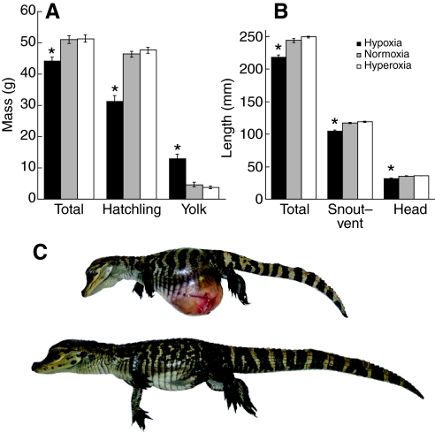
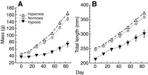
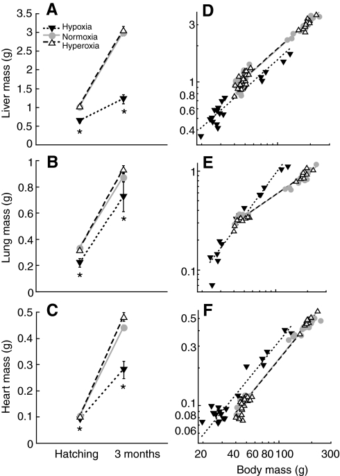
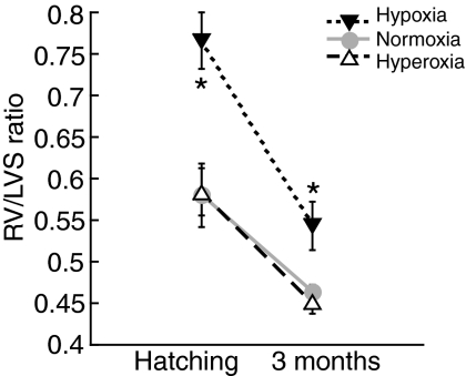
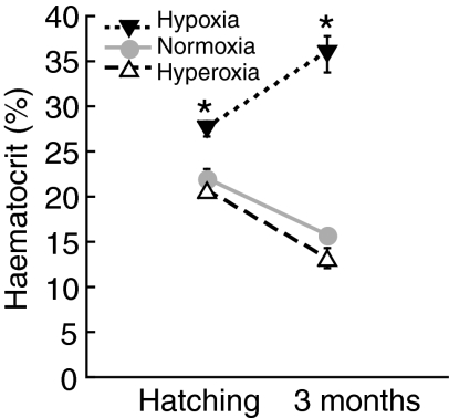
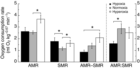
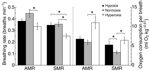
Recent palaeoatmospheric models suggest large-scale fluctuations in ambient oxygen level over the past 550 million years. To better understand how global hypoxia and hyperoxia might have affected the growth and physiology of contemporary vertebrates, we incubated eggs and raised hatchlings of the American alligator. Crocodilians are one of few vertebrate taxa that survived these global changes with distinctly conservative morphology. We maintained animals at 30 degrees C under chronic hypoxia (12% O(2)), normoxia (21% O(2)) or hyperoxia (30% O(2)). At hatching, hypoxic animals were significantly smaller than their normoxic and hyperoxic siblings. Over the course of 3 months, post-hatching growth was fastest under hyperoxia and slowest under hypoxia. Hypoxia, but not hyperoxia, caused distinct scaling of major visceral organs-reduction of liver mass, enlargement of the heart and accelerated growth of lungs. When absorptive and post-absorptive metabolic rates were measured in juvenile alligators, the increase in oxygen consumption rate due to digestion/absorption of food was greatest in hyperoxic alligators and smallest in hypoxic ones. Hyperoxic alligators exhibited the lowest breathing rate and highest oxygen consumption per breath. We suggest that, despite compensatory cardiopulmonary remodelling, growth of hypoxic alligators is constrained by low atmospheric oxygen supply, which may limit their food utilisation capacity. Conversely, the combination of elevated metabolism and low cost of breathing in hyperoxic alligators allows for a greater proportion of metabolised energy to be available for growth. This suggests that growth and metabolic patterns of extinct vertebrates would have been significantly affected by changes in the atmospheric oxygen level.


Figures

Fig. 1.
Fig. 1.
Comparison of alligator hatchlings incubated under three different oxygen levels (12%, 21% and 30%). (A) Mass measurements: total, body and yolk masses. Hypoxic hatchlings are significantly smaller than their normoxic and hyperoxic siblings, but the remaining yolk sac of hypoxic animals is significantly larger. (B) Length measurements: total, snout-to-vent and head lengths. Hypoxic hatchlings are significantly smaller than their normoxic and hyperoxic siblings. (C) A pair of anaesthetised alligator siblings, incubated under hypoxia (above) and normoxia (below). Note the diminutive hatchling size and the protruding yolk sac in the hypoxic animal. The yolk sac is completely incorporated into the abdominal cavity and the umbilical scar closed, but the abdominal skin is stretched thin and a pronounced left umbilical vein is seen. The height of the yolk sac exceeds the length of the limbs, making locomotion cumbersome. Statistical significance between groups was calculated by ANOVA with post hoc Tukey–Kramer (*P<0.05). Bar height and error bars indicate the mean ± s.e.m. for each group.
Fig. 2.
Fig. 2.
Growth curves of alligator juveniles under three oxygen levels (hypoxia, normoxia and hyperoxia) over 3 months post-hatching. (A) Body mass and (B) total length growth of alligators. Hypoxic animals grew slowest, and hyperoxic animals grew fastest in terms of body mass and total length. Symbols with error bars indicate the mean ± s.e.m. for each group.
Fig. 3.
Fig. 3.
(A–C) Absolute wet masses of major visceral organs at hatching and 3 months later: (A) liver, (B) lungs and (C) heart. All organs are significantly smaller in hypoxic alligators at both ages (ANOVA with post hoc Tukey–Kramer test, *P<0.05). Symbols and error bars indicate the mean ± s.e.m. (D–F) Ontogenetic allometry of major visceral organs in alligators reared under hypoxia, normoxia and hyperoxia. (D) Liver scales to Mb0.80 (Mb, body mass) in all groups, but is significantly smaller in hypoxic animals. (E) Lungs scale similarly (Mb0.73) in normoxia and hyperoxia, but exhibit a significantly steeper slope (Mb1.44) in the hypoxic group. (F) Heart scales with slight positive allometry (Mb1.07) in all groups, but is significantly larger in hypoxic alligators.
Fig. 4.
Fig. 4.
RV/LVS ratio of the right ventricle (free wall only) to left ventricle (free wall and interventricular septum) in hatchling and juvenile alligators. In both age groups, the ratio is significantly higher in hypoxic animals than in either their normoxic or hyperoxic siblings, but no significant difference exists between the last two groups. Statistical significance between groups was calculated by Wilcoxon/Kruskal–Wallis rank sums test with post hoc Tukey–Kramer (*P<0.05).
Fig. 5.
Fig. 5.
Haematocrit levels in hatchling and juvenile alligators reared under chronic hypoxia, normoxia and hyperoxia. Haematocrit level is significantly higher in hypoxic animals, but not significantly different between normoxic and hyperoxic animals. Statistical significance between groups was calculated by Wilcoxon/Kruskal–Wallis rank sums test with post hoc Tukey–Kramer (*P<0.05).
Fig. 6.
Fig. 6.
Differences in metabolic rate of juvenile alligators in different oxygen atmospheres under absorptive and post-absorptive (standard) conditions. Absorptive (AMR) and standard (SMR) metabolic rates are expressed as mass-corrected oxygen consumption rate. Absolute (AMR–SMR) and relative (AMR:SMR) metabolic elevation due to digestion/absorption are also plotted. Metabolic rates and absolute metabolic elevation are highest in hyperoxic alligators. Statistical significance between groups was calculated by ANOVA with post hoc Tukey–Kramer (*P<0.05). Bar height and error bars indicate the mean ± s.e.m.
Fig. 7.
Fig. 7.
Breathing rate (left) and mass-corrected oxygen consumption per breath (right) of 3 month old alligators in different oxygen atmospheres under absorptive and post-absorptive (standard) conditions. Hyperoxic alligators show significantly lower breathing rates than their siblings in normoxic and hypoxic groups. Conversely, each breath supports greater oxygen consumption in hyperoxic animals than in other groups. Statistical significance between groups was calculated by ANOVA with post hoc Tukey–Kramer (*P<0.05). Bar height and error bars indicate the mean ± s.e.m.

Similar articles

Cited by

References

    1. Andrade, D. V. and Abe, A. S. (1999). Gas exchange and ventilation during dormancy in the tegu lizard Tupinambis merianae. J. Exp. Biol. 202, 3677-3685. - PubMed
    1. Andrews, R. M. (2001). Low oxygen: a constraint on the evolution of viviparity in reptiles. Physiol. Biochem. Zool. 75, 145-154. - PubMed
    1. Bartlett, D., Jr and Remmers, J. E. (1971). Effects of high altitude exposure on the lungs of young rats. Respir. Physiol. 13, 116-125. - PubMed
    1. Bavis, R. W. (2005). Developmental plasticity of the hypoxic ventilatory response after perinatal hyperoxia and hypoxia. Respir. Physiol. Neurobiol. 149, 287-299. - PubMed
    1. Bergman, N. M., Lenton, T. M. and Watson, A. J. (2004). COPSE: a new model of biogeochemical cycling over Phanerozoic time. Am. J. Sci. 304, 397-437.

Publication types