. 2009 May;12(5):568-76.
doi: 10.1038/nn.2318. Epub 2009 Apr 19.

Myosin-dependent targeting of transmembrane proteins to neuronal dendrites

Affiliations

Myosin-dependent targeting of transmembrane proteins to neuronal dendrites

Tommy L Lewis Jr et al. Nat Neurosci. 2009 May.

Abstract

The distinct electrical properties of axonal and dendritic membranes are largely a result of specific transport of vesicle-bound membrane proteins to each compartment. How this specificity arises is unclear because kinesin motors that transport vesicles cannot autonomously distinguish dendritically projecting microtubules from those projecting axonally. We hypothesized that interaction with a second motor might enable vesicles containing dendritic proteins to preferentially associate with dendritically projecting microtubules and avoid those that project to the axon. Here we show that in rat cortical neurons, localization of several distinct transmembrane proteins to dendrites is dependent on specific myosin motors and an intact actin network. Moreover, fusion with a myosin-binding domain from Melanophilin targeted Channelrhodopsin-2 specifically to the somatodendritic compartment of neurons in mice in vivo. Together, our results suggest that dendritic transmembrane proteins direct the vesicles in which they are transported to avoid the axonal compartment through interaction with myosin motors.


Figures
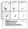
Figure 1
Figure 1
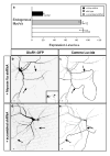
Myosin function is necessary for targeting of exogenous dendritic transmembrane proteins. (ac) In a cortical neuron expressing coexpressing HA-mCherry (a), GluR1-GFP (b) localized specifically to the somatodendritic region. (c) Camera lucida drawing Axon, black; somatodendritic region, gray. (df) In contrast, when expressed with a dominant negative variant of Myosin Va (HA-dnMVa) (d), GluR1-GFP (e) localized nonspecifically. (f) Camera lucida drawing. (g) The axon-to-dendrite ratios (ADRs; see Online Methods) of GluR1-GFP, Kv4.2-MYC and EAAT3-HA expressed with tagged dnMVa were at least fourfold higher than when each was expressed with either HA-mCherry or GFP: ADRGluR1,dnMVa = 0.97 ± 0.17; ADRGluR1,mCherry = 0.16 ± 0.02; ADRKV4.2,dnMVa = 0.86 ± 0.19; ADRKv4.2,HA-mCherry = 0.17 ± 0.03; ADREAAT3-HA,dnMVa = 1.27 ± 0.17; ADREAAT3-HA,HA-mCherry = 0.30 ± 0.05. Conversely, the ADR of GluR1-GFP was not significantly different when it is expressed with HA-dnMVb (ADRGluR1,dnMVb = 0.25 ± 0.03) from the ADR when it was expressed with HA-mCherry. Error bars, s.e.m.; arrow, axon; arrowheads, axon initial segment. Insets: staining for Ankyrin G, used to identify the axon. Scale bars, 10 μm. *P < 0.0002; NS, P > 0.08 (Wilcoxon-Mann-Whitney).
Figure 2
Figure 2
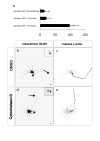
Myosin function is necessary for targeting of endogenous dendritic transmembrane proteins. (ac) In a cortical neuron expressing GFP (a), endogenous EAAT3 (b) localized preferentially to the somatodendritic region. (c) Camera lucida drawing. Axon, black; somatodendritic region, gray. (df) In contrast, when expressed with a dominant negative variant of Myosin Va (d), endogenous EAAT3 (e) localized to both the somatodendritic and axonal compartments. (f) Camera lucida drawing. (g) The ADRs of endogenous GluR1 and EAAT3 expressed with tagged dnMVa were approximately threefold higher than when each was expressed with either HA-mCherry or GFP: ADRGluR1,dnMVa = 1.03 ± 0.13; ADRGluR1,GFP = 0.31 ± 0.05; ADREAAT3,dnMVa = 0.92 ± 0.13; ADREAAT3,GFP = 0.32 ± 0.04. Error bars, s.e.m.; arrow, axon; arrowheads, axon initial segments. Insets, staining for Ankyrin G. Scale bars, 10 μm; *P < 0.0009 (Wilcoxon-Mann-Whitney).
Figure 3
Figure 3
Knockdown of Myosin Va expression blocks dendritic targeting of GluR1-GFP. (a) Expression of siRNA directed against Myosin Va in cortical neurons in dissociated culture caused at least a 75% drop in the expression of endogenous Myosin Va (P < 0.0001; a.u., arbitrary units). (b) Expression of MVa siRNA with GluR1-GFP caused the receptor to localize nonspecifically. (c) Camera lucida drawing of neuron shown in b. Axon, black; somatodendritic region, gray. (d) GluR1-GFP localized to dendrites when expressed with a scrambled siRNA. Inset, staining of endogenous Myosin Va in the neuron shown in b (arrowhead) and in an untransfected control. (e) Camera lucida drawing of neuron shown in d. Error bars, s.e.m.; arrow, axon. The ADR is unnormalized (see Online Methods), so that only the relative values are relevant. Axons were identified by either the presence of Ankyrin G or the absence of MAP2. Scale bars, 10 μm; *P < 0.001 (Wilcoxon-Mann-Whitney).
Figure 4
Figure 4
Association with Myosin Va is sufficient for dendritic targeting. (a c) CD8 fused to a Myosin Va binding domain (CD8-MBD) and expressed with GFP in a cortical neuron localized specifically to the soma and dendrites. (c) Camera lucida drawing. Axon, black; somatodendritic region, gray. (df) CD8 expressed with GFP localized to both axons and dendrites. (f) Camera lucida drawing. (g) CD8-MBD, which associates with Myosin Va, localized dendritically as assessed by either total protein or surface protein staining (ADRCD8-MBD = 0.13 ± 0.02; ADRCD8-MBD,surface = 0.19 ± 0.04). In contrast, CD8 localized nonspecifically in either case (ADRCD8 = 0.90 ± 0.08; ADRCD8,surface = 0.80 ± 0.13). CD8-MBD localized nonspecifically when expressed with HA-dnMVa (ADRCD8-MBD,dnMVa = 1.14 ± 0.23). Error bars, s.e.m.; arrow, axon; arrowheads, axon initial segment. Insets, staining for Ankyrin G. Scale bars, 10 μm. *P < 0.0002; NS, P > 0.08 (Wilcoxon-Mann-Whitney).
Figure 5
Figure 5
Role of actin in dendritic localization. (ac) In a cortical neuron exposed to cytochalasin D for approximately 20 h, both HA-mCherry (a) and GluR1-GFP (b) localized nonspecifically. (c) Camera lucida drawing. Axon, black; somatodendritic compartment, gray. (df) In contrast, in cortical neurons exposed to DMSO, GluR1-GFP expression (e) was enriched in the dendrites. (f) Camera lucida drawing. (g) ADRs confirm that when exogenous dendritic proteins were expressed in cortical neurons exposed to cytochalasin D, they localized nonspecifically (ADRTfR = 1.01 ± 0.09; ADREAAT3 = 0.87 ± 0.09; ADRNlg = 1.07 ± 0.19; ADRKv4.2 = 0.76 ± 0.13; ADRGluR1 = 0.91 ± 0.12), whereas when the same proteins were expressed in similar neurons exposed to DMSO alone, they localized specifically to dendrites (ADRTfR = 0.20 ± 0.03; ADREAAT3 = 0.15 ± 0.03; ADRNlg = 0.14 ± 0.03; ADRKv4.2 = 0.14 ± 0.03; ADRGlur1 = 0.10 ± 0.02). *P < 0.0002 (Wilcoxon-Mann-Whitney); error bars, s.e.m.; arrow, axon; arrowheads, axon initial segment. Insets, staining for Ankyrin G. Scale bars, 10 μm.
Figure 6
Figure 6
Role of actin filaments in the initial targeting of GluR1 to dendrites. (a) In the presence of thrombin, GFP was efficiently cleaved from GluR1-TCS-GFP, which effectively removes the label from all protein that is present on the cell surface, allowing specific labeling of intracellular protein that has never been to the cell surface. The surface staining of GluR1 in the presence of thrombin can largely be attributed to background, as it is similar to the staining seen in neurons that were not transfected. (b,c) In a cortical neuron exposed to DMSO and thrombin, intracellular GluR1 (b) localized specifically to the somatodendritic compartment. (c) Camera lucida drawing. Axon, black; somatodendritic compartment, gray. (d,e) However, in a cell exposed to cytochalasin D and thrombin, intracellular protein localized to both axons and dendrites. Error bars, s.e.m.; Arrow, axon; arrowheads, axon initial segment. Insets, staining for Ankyrin G. Scale bars, 10 μm.
Figure 7
Figure 7
Targeting ChR2 to dendrites of pyramidal neurons using a myosin-binding domain. (a) Coronal section through the somatosensory cortex from mice expressing ChR2 (left) and ChR2-MBD (right) tagged with fluorescent proteins in L2/3 pyramidal neurons. Cortical layers are indicated at left (b) Fluorescence intensity (Norm. intensity), normalized to the maximum for each slice, as a function of cortical depth. (c) Confocal images of L2/3 in mice showing expression of ChR2 (left) and ChR2-MBD (right) in green. Blue, DAPI stain showing nuclei. (d) Enlargements of regions boxed in c. (e) Image of a brain slice. Blue box, photostimulation area. Green triangle, position of soma. (f) Left, schematic of a L2/3 cell and the photostimulation pattern (16 × 8, 50-μm spacing). Right, traces, recorded in current clamp, corresponding to dashed box on left. (g) Excitation profiles of layer 2/3 neurons expressing ChR2 (top) and ChR2-MBD (bottom) at different laser powers (size of stimulation area corresponds to blue box in e). AP, action potentials The ChR2-MBD map at 8 μW corresponds to the data in f. (h) The number of action potentials in L5 versus L2/3. Data are pooled across cells (ChR2, n = 9; ChR2-MBD, n = 15) and powers (5–350 μW). Slopes of the regression lines are 0.8 (ChR2) and 0.13 (ChR2-MBD) (P < 0.001). In some instances, several points with the same value are plotted on top of each other (Supplementary Fig. 11).

Similar articles

Cited by

References

    1. Mostov K, Su T, ter Beest M. Polarized epithelial membrane traffic: conservation and plasticity. Nat Cell Biol. 2003;5:287–293. - PubMed
    1. Folsch H, Ohno H, Bonifacino JS, Mellman I. A novel clathrin adaptor complex mediates basolateral targeting in polarized epithelial cells. Cell. 1999;99:189–198. - PubMed
    1. Burack MA, Silverman MA, Banker G. The role of selective transport in neuronal protein sorting. Neuron. 2000;26:465–472. - PubMed
    1. Matsuda S, et al. Accumulation of AMPA receptors in autophagosomes in neuronal axons lacking adaptor protein AP-4. Neuron. 2008;57:730–745. - PubMed
    1. Setou M, et al. Glutamate-receptor-interacting protein GRIP1 directly steers kinesin to dendrites. Nature. 2002;417:83–87. - PubMed

Publication types

MeSH terms