Phasic acetylcholine release and the volume transmission hypothesis: time to move on
- PMID: 19377503
- PMCID: PMC2699581
- DOI: 10.1038/nrn2635
Phasic acetylcholine release and the volume transmission hypothesis: time to move on
Abstract
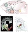
Traditional descriptions of the cortical cholinergic input system focused on the diffuse organization of cholinergic projections and the hypothesis that slowly changing levels of extracellular acetylcholine (ACh) mediate different arousal states. The ability of ACh to reach the extrasynaptic space (volume neurotransmission), as opposed to remaining confined to the synaptic cleft (wired neurotransmission), has been considered an integral component of this conceptualization. Recent studies demonstrated that phasic release of ACh, at the scale of seconds, mediates precisely defined cognitive operations. This characteristic of cholinergic neurotransmission is proposed to be of primary importance for understanding cholinergic function and developing treatments for cognitive disorders that result from abnormal cholinergic neurotransmission.
Figures


Similar articles
-
Forebrain Cholinergic Signaling: Wired and Phasic, Not Tonic, and Causing Behavior.J Neurosci. 2020 Jan 22;40(4):712-719. doi: 10.1523/JNEUROSCI.1305-19.2019. J Neurosci. 2020. PMID: 31969489 Free PMC article. Review.
-
Nicotinic Transmission onto Layer 6 Cortical Neurons Relies on Synaptic Activation of Non-α7 Receptors.Cereb Cortex. 2016 Jun;26(6):2549-2562. doi: 10.1093/cercor/bhv085. Epub 2015 May 1. Cereb Cortex. 2016. PMID: 25934969
-
Synaptic Release of Acetylcholine Rapidly Suppresses Cortical Activity by Recruiting Muscarinic Receptors in Layer 4.J Neurosci. 2018 Jun 6;38(23):5338-5350. doi: 10.1523/JNEUROSCI.0566-18.2018. Epub 2018 May 8. J Neurosci. 2018. PMID: 29739869 Free PMC article.
-
Deterministic functions of cortical acetylcholine.Eur J Neurosci. 2014 Jun;39(11):1912-20. doi: 10.1111/ejn.12515. Epub 2014 Mar 4. Eur J Neurosci. 2014. PMID: 24593677 Free PMC article. Review.
-
Regulation of cortical acetylcholine release: insights from in vivo microdialysis studies.Behav Brain Res. 2011 Aug 10;221(2):527-36. doi: 10.1016/j.bbr.2010.02.022. Epub 2010 Feb 16. Behav Brain Res. 2011. PMID: 20170686 Free PMC article. Review.
Cited by
-
Rapid multi-directed cholinergic transmission in the central nervous system.Nat Commun. 2021 Mar 2;12(1):1374. doi: 10.1038/s41467-021-21680-9. Nat Commun. 2021. PMID: 33654091 Free PMC article.
-
Novel interplay between agonist and calcium binding sites modulates drug potentiation of α7 acetylcholine receptor.Cell Mol Life Sci. 2024 Aug 7;81(1):332. doi: 10.1007/s00018-024-05374-1. Cell Mol Life Sci. 2024. PMID: 39110172 Free PMC article.
-
Basal Forebrain Mediates Motivational Recruitment of Attention by Reward-Associated Cues.Front Neurosci. 2018 Oct 30;12:786. doi: 10.3389/fnins.2018.00786. eCollection 2018. Front Neurosci. 2018. PMID: 30425617 Free PMC article.
-
Fast modulation of visual perception by basal forebrain cholinergic neurons.Nat Neurosci. 2013 Dec;16(12):1857-1863. doi: 10.1038/nn.3552. Epub 2013 Oct 27. Nat Neurosci. 2013. PMID: 24162654 Free PMC article.
-
Forebrain Cholinergic Signaling: Wired and Phasic, Not Tonic, and Causing Behavior.J Neurosci. 2020 Jan 22;40(4):712-719. doi: 10.1523/JNEUROSCI.1305-19.2019. J Neurosci. 2020. PMID: 31969489 Free PMC article. Review.
References
-
- Everitt BJ, Robbins TW. Central cholinergic systems and cognition. Annu Rev Psychol. 1997;48:649–684. - PubMed
-
- Sarter M, Gehring WJ, Kozak R. More attention must be paid: the neurobiology of attentional effort. Brain Res Rev. 2006;51:145–160. - PubMed
-
- Sarter M, Givens B, Bruno JP. The cognitive neuroscience of sustained attention: where top-down meets bottom-up. Brain Res Rev. 2001;35:146–160. - PubMed
-
- Sarter M, Hasselmo ME, Bruno JP, Givens B. Unraveling the attentional functions of cortical cholinergic inputs: interactions between signal-driven and top-down cholinergic modulation of signal detection. Brain Res Rev. 2005;48:98–111. - PubMed
Publication types
MeSH terms
Substances
Grants and funding
LinkOut - more resources
Full Text Sources

