

Review
doi: 10.1038/nrc2608.
Epub 2009 Apr 20.
DNA topoisomerase II and its growing repertoire of biological functions
Affiliations
- PMID: 19377505
- PMCID: PMC2730144
- DOI: 10.1038/nrc2608
Item in Clipboard
Review
DNA topoisomerase II and its growing repertoire of biological functions
Nat Rev Cancer.
2009 May.
Abstract
DNA topoisomerases are enzymes that disentangle the topological problems that arise in double-stranded DNA. Many of these can be solved by the generation of either single or double strand breaks. However, where there is a clear requirement to alter DNA topology by introducing transient double strand breaks, only DNA topoisomerase II (TOP2) can carry out this reaction. Extensive biochemical and structural studies have provided detailed models of how TOP2 alters DNA structure, and recent molecular studies have greatly expanded knowledge of the biological contexts in which TOP2 functions, such as DNA replication, transcription and chromosome segregation -- processes that are essential for preventing tumorigenesis.
Figures
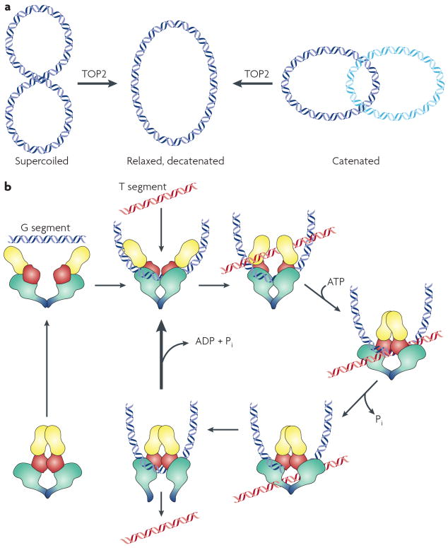
A. Reactions catalyzed by eukaryotic Top2 include decatenation of linked intact double stranded DNA and relaxation of supercoiled DNA. The reaction formally requires introduction of a double strand break, strand passage, and break resealing. B. Topoisomerase II interacts with two DNA strands to effect strand passage. The enzyme introduces a double strand break in one DNA strand, termed the G or “gate segment”, and will pass a second strand termed the T segment through the break. In the presence of Mg2+, the enzyme can cleave the DNA, forming a phosphotyrosine linkage between each single strand and a tyrosine in each subunit. ATP binding causes the enzyme to form a closed clamp. The closed clamp may also capture another strand (the T strand) that will pass through the break made in the G strand. After passing through the break in the G strand, the T strand exits the enzyme through the carboxy terminus (the bottom of the enzyme as drawn). ATP hydrolysis occurs at two steps in the reaction cycle. The first ATP hydrolyzed may assist in strand passage. The second hydrolysis step (along with release of ADP and Pi) allows the clamp to re-open, and allows release of the G segment (for a distributive reaction). Alternately, the enzyme may initiate another catalytic cycle without dissociating from the G strand.
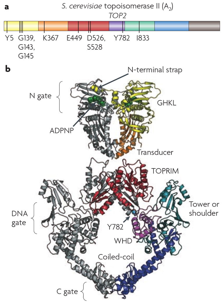
A. Domain structure of a eukaryotic Top2. Domains are indicated in color, and key residues are indicated. The residues marked include G139, G143 and G145 in the ATP binding domain, Lys367, a transducer domain residue that contributes to the ATPase; Glu449, Asp526, and Asp 528 the acidic triad involved in binding a divalent cation; Tyr782, the residue that makes a covalent attachment with DNA, and Ile833, a tower domain residue that is involved in DNA interaction. B. Structure of yeast Top2 based on structures for the ATPase domain and the breakage reunion domain,. The GHKL and transducer domain is shown in yellow and orange, TOPRIM, winged helix, tower, and coiled coil are shown in red, purple, teal and blue, and Tyr782 is shown as a cyan sphere. The figure is from James Berger .
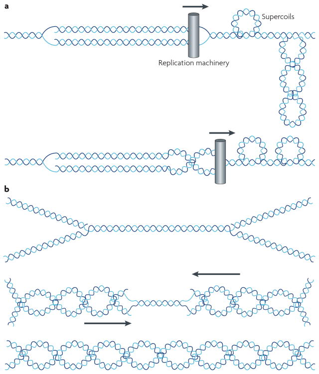
A. Partition of superhelical strain during replication fork progression in vivo. During replication helicase action on DNA creates positive superhelical stress on the DNA. This results in positive supercoils in front of the fork, as shown in A. The structure shown in A can isomerize into intertwinings of the daughter duplexes, generating precatenanes, as shown in B. (Figure is from Postow and Cozzarelli, Bioessays 21:805, 1999). B. At early steps in replication, when forks are widely separated, either Top1 or Top2 can function as a replication swivel. Top1 acts by relaxing positive supercoils while Top2 unlinks precatenanes. Note that Top2 also should be able to relax positive supercoils, and does not require the isomerization to precatenanes for unlinking replicated strands. As the replication forks converge, there is a limited ability to generate positive supercoiling, and complete unlinking absolutely requires Top2.
References
-
- Champoux JJ. DNA topoisomerases: structure, function, and mechanism. Annu Rev Biochem. 2001;70:369–413. - PubMed
-
- Wang JC. Moving one DNA double helix through another by a type II DNA topoisomerase: the story of a simple molecular machine. Q Rev Biophys. 1998;31:107–44. - PubMed
-
- Sundin O, Varshavsky A. Arrest of segregation leads to accumulation of highly intertwined catenated dimers: dissection of the final stages of SV40 DNA replication. Cell. 1981;25:659–69. - PubMed
-
- Sundin O, Varshavsky A. Terminal stages of SV40 DNA replication proceed via multiply intertwined catenated dimers. Cell. 1980;21:103–14. - PubMed
-
- Bates AD, Maxwell A. DNA topology. Oxford University Press; Oxford: 2005.
Publication types
MeSH terms
Substances
Grants and funding
LinkOut - more resources
Full Text Sources
Other Literature Sources
Miscellaneous
