. 2009 May 28;28(21):2163-72.
doi: 10.1038/onc.2009.82. Epub 2009 Apr 20.

Wnt pathway aberrations including autocrine Wnt activation occur at high frequency in human non-small-cell lung carcinoma

Affiliations

Wnt pathway aberrations including autocrine Wnt activation occur at high frequency in human non-small-cell lung carcinoma

G Akiri et al. Oncogene. .

Abstract

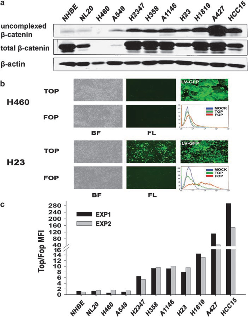
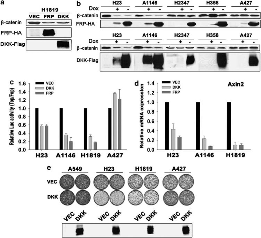
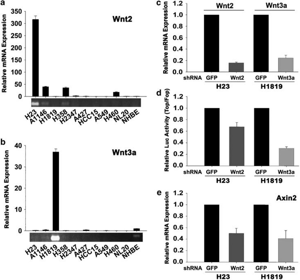
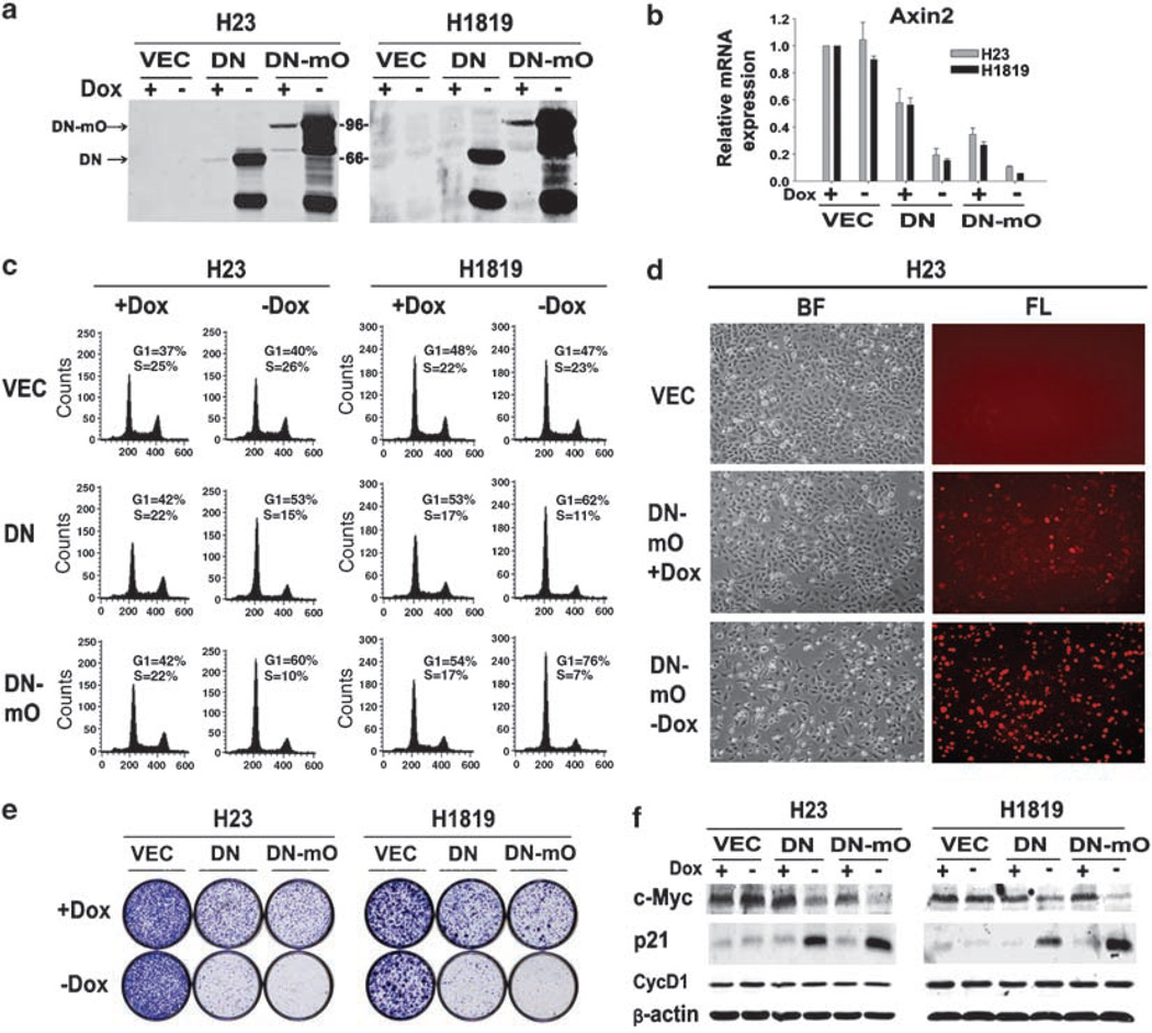
Lung cancer is the most common cause of cancer mortality worldwide. Non-small-cell lung carcinomas (NSCLCs), which represent around 80% of lung tumors, exhibit poor prognosis and are usually refractory to conventional chemotherapy. Elucidating the molecular and cellular mechanisms that are dysregulated in NSCLCs may lead to new possibilities for targeted therapy or enhanced efficacy of current therapies. Here we demonstrate Wnt pathway activation in around 50% of human NSCLC cell lines and primary tumors, through different mechanisms, including autocrine Wnt pathway activation involving upregulation of specific Wnt ligands. Downregulation of activated Wnt signaling inhibited NSCLC proliferation and induced a more differentiated phenotype. Together, our findings establish importance of activated Wnt signaling in human NSCLCs and offer the possibility of targeting upregulated Wnt signaling as a new therapeutic modality for this disease.


Conflict of interest statement

Conflict of interest

The authors declare no conflict of interest.

Figures

Figure 1
Figure 1
Wnt signaling activation in human non-small-cell lung carcinoma (NSCLC) cell lines. (a) Total cell lysates (1 mg) were subjected to precipitation with a glutathione S-transferase (GST)-E cadherin fusion protein (Bafico et al., 1998). Total cell lysates (25 µg) and the GST-E-cadherin precipitates were subjected to immunoblot analysis with an mAb directed against β-catenin. (b) Fluorescence-activated cell sorting (FACS) analysis, phase contrast and fluorescence images of H460 (upper panel) and H23 (lower panel) NSCLC cells infected with TOP or FOP TCF-GFP reporter lentiviruses or with enhanced green fluorescent protein (EGFP) expressing lentivirus (LV-GFP). BF, bright field; FL, fluorescence. (c) Lentivirus-mediated TCF-GFP reporter activity in human NSCLC cells. Results are depicted as the ratio TOP/FOP GFP mean fluorescence intensity (MFI). Results from two independent experiments are shown.
Figure 2
Figure 2
Effects of FRP1 and DKK1 inhibition on Wnt/β-catenin signaling and growth of human non-small-cell lung carcinoma (NSCLC) cells. (a) Effects of constitutive expression of FRP1 and DKK1 on uncomplexed β-catenin in H1819 NSCLC cell line. FRP1 and DKK1 expression was determined by immunoblot analysis as described in Materials and methods. (b) Analysis of NSCLC lines for uncomplexed β-catenin using regulatable expression of hemagglutinin (HA)-tagged FRP1 (upper panel) and Flag-tagged DKK1 (lower panel). NSCLC cells expressing Tet regulatable FRP1-HA or DKK1-Flag were generated as described in Supplementary materials and methods. Expression of FRP1-HA or DKK1-Flag was induced by removal of dox from the culture medium. Cells expressing tetracycline Trans-activator (tTa) were used as control. Analysis of uncomplexed β-catenin was performed as described in Materials and methods using 1mg total cell lysates, except for A427 cells, where 0.1 mg cell lysate was used. FRP1 and DKK1 expression was determined by immunoblot analysis as described in Materials and methods. (c) FRP1- and DKK1-mediated inhibition of TCF luciferase reporter activity in NSCLC cell lines. Luciferase reporter activity was calculated by dividing the TOP/RL ratio by the FOP/RL ratio. Results were normalized to the results with vector-transduced cultures. The values represent the mean ± s.d. from two independent experiments. (d) Real-time PCR quantification of FRP1 and DKK1 effects on axin2 mRNA expression. H23, A1146 and H1819 cells were infected with vector (VEC), FRP1-HA or DKK1-Flag lentiviruses. qRT–PCR was performed as described in Supplementary materials and methods. Relative mRNA expression levels were quantified using the ΔΔC(t) method (Pfaffl, 2001). (e) Effects of DKK1 on cell growth. A549, H23, H1819 and A427 cells were infected with lentiviruses expressing vector (VEC) or DKK1-Flag and 2 × 10⁴ cells were plated into 60mm tissue culture dishes. Cultures were visualized using crystal violet staining 2–3 weeks after plating. Expression of Flag-tagged DKK1 was assessed by immunoblot analysis as described in Materials and methods.
Figure 3
Figure 3
Overexpression of Wnt2 and Wnt3a contributes to constitutive Wnt activation in autocrine non-small-cell lung carcinoma (NSCLC) cells. (a, b) Real-time PCR quantification of Wnt2 (a) and Wnt3a (b) expression in H23 and H1819 cells, respectively. To visualize relative expression levels of Wnt2 and Wnt3a, qPCR reactions were removed before saturation and PCR products were separated on 1.5% agarose gel and stained with ethidium bromide. (c) shRNA knockdown quantification of Wnt2 and Wnt3a. H23 and H1819 cells were infected with lentiviruses expressing shRNA targeting green fluorescent protein (GFP), Wnt2 or Wnt3a. (d) Effect of shRNA knockdown of Wnt2 and Wnt3a on TCF reporter activity. Luciferase reporter activity was calculated by dividing the TOP/RL ratio by FOP/RL ratio. Each column represents the mean ± s.d. of two independent experiments. (e) Effect of shRNA knockdown of Wnt2 and Wnt3a on axin2 mRNA expression. H23 and H1819 cells were infected with lentiviruses expressing shRNA targeting GFP, Wnt2 or Wnt3a.
Figure 4
Figure 4
Effects of inducible dominant-negative TCF-4 (DNTCF-4) on growth of non-small-cell lung carcinoma (NSCLC) autocrine cells. (a) Immunoblot analysis of DNTCFs expression. H23 and H1819 cells were infected with lentiviruses expressing DNTCF-4 (DN), DN-mOrange (DN-mO) and vector (VEC) under the control of a tetracycline-inducible promoter and selected with puromycin in the presence of dox. After washing, cells were divided into separate cell culture dishes in the presence or absence of dox and analysed by immunoblot 3 days after induction. Expression of DNTCF-4 proteins was detected using an antibody to TCF-4. Lower molecular weight immunoreactive DNTCF species were also observed. Molecular weights in kilodaltons are indicated (kDa). (b) Effect of DNTCFs on axin2 mRNA expression. RNA was extracted from H23 and H1819 cells infected as in (a) and maintained in the presence or absence of dox for 3 days. (c) Effect of DNTCFs on cell-cycle profile. Propidium iodide (PI) analysis of H23 and H1819 cells infected as in (a) and maintained in the presence or absence of dox at 3 days after induction. Numbers indicate the percentage of cells in G1 or S phase for each cell line analysed. Results are representative of at least two independent experiments. (d) Effects on proliferation of H23 cells expressing inducible DN-mO in the presence or absence of dox, observed at 3 days after induction. BF, bright field; FL, fluorescence. (e) Effects on growth of H23 and H1819 at 2–3 weeks following expression of VEC, DN and DN-mO. A total of 2 × 10⁴ cells were plated into 60mm plates in the presence or absence of dox. Cultures were visualized by crystal violet staining. (f) Effects of DNTCFs on expression of c-Myc, cyclin D1 and p21. Protein lysates from H23 and H1819 cells, infected with VEC, DN and DN-mO, and grown in the presence or absence of dox for 3 days, were analysed by immunoblot. Dox, doxycycline.

Similar articles

Cited by

References

    1. Bafico A, Gazit A, Wu-Morgan SS, Yaniv A, Aaronson SA. Characterization of Wnt-1 and Wnt-2 induced growth alterations and signaling pathways in NIH3T3 fibroblasts. Oncogene. 1998;16:2819–2825. - PubMed
    1. Bafico A, Liu G, Goldin L, Harris V, Aaronson SA. An autocrine mechanism for constitutive Wnt pathway activation in human cancer cells. Cancer Cell. 2004;6:497–506. - PubMed
    1. Bernasconi NL, Wormhoudt TAM, Laird-Offringa IA. Post-transcriptional deregulation of myc genes in lung cancer cell lines. Am J Respir Cell Mol Biol. 2000;23:560–565. - PubMed
    1. Braga VM, Pemberton LF, Duhig T, Gendler SJ. Spatial and temporal expression of an epithelial mucin, Muc-1, during mouse development. Development. 1992;115:427–437. - PubMed
    1. Clevers H. Wnt/beta-catenin signaling in development and disease. Cell. 2006;127:469–480. - PubMed

Publication types

MeSH terms

Substances