. 2009 Apr 17;137(2):332-43.
doi: 10.1016/j.cell.2009.02.018.

Platelet matrix metalloprotease-1 mediates thrombogenesis by activating PAR1 at a cryptic ligand site

Affiliations

Platelet matrix metalloprotease-1 mediates thrombogenesis by activating PAR1 at a cryptic ligand site

Vishal Trivedi et al. Cell. .

Abstract

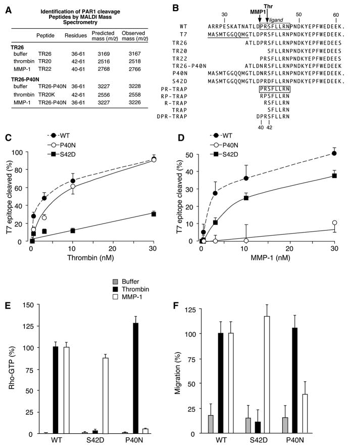
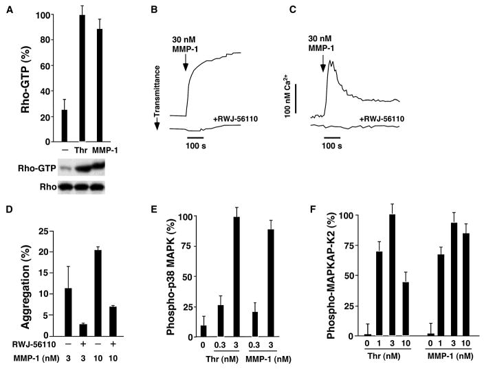
Matrix metalloproteases (MMPs) play important roles in normal and pathological remodeling processes including atherothrombotic disease, inflammation, angiogenesis, and cancer. MMPs have been viewed as matrix-degrading enzymes, but recent studies have shown that they possess direct signaling capabilities. Platelets harbor several MMPs that modulate hemostatic function and platelet survival; however their mode of action remains unknown. We show that platelet MMP-1 activates protease-activated receptor-1 (PAR1) on the surface of platelets. Exposure of platelets to fibrillar collagen converts the surface-bound proMMP-1 zymogen to active MMP-1, which promotes aggregation through PAR1. Unexpectedly, MMP-1 cleaves PAR1 at a distinct site that strongly activates Rho-GTP pathways, cell shape change and motility, and MAPK signaling. Blockade of MMP1-PAR1 curtails thrombogenesis under arterial flow conditions and inhibits thrombosis in animals. These studies provide a link between matrix-dependent activation of metalloproteases and platelet-G protein signaling and identify MMP1-PAR1 as a potential target for the prevention of arterial thrombosis.


Figures

Figure 1
Figure 1. Collagen Generates Active MMP-1 on Platelets Which Cleaves the PAR1 N-terminal Extracellular Domain
(A) Human platelets (106/μL) were treated for 15 min with either 20 μM ADP, 20 μM U-46619, or 20 μg/ml type-I collagen. The enzymatic activity of active MMP-1 in supernatants and platelet lysates was determined using DQ collagen I as fluorogenic substrate in the presence or absence of 3 μM FN-439, or 20 μg/ml each of MMP1-blocking Ab or control IgG, with APMA-activated MMP-1 serving as control (striped bars). (B) Pellets and supernatants were collected from platelets (250,000/μL) stimulated with the agonists in (A) or convulxin (1 μg/ml). Concentrations of released and platelet-associated MMP-1 pro-domains were measured by ELISA. (C) Surface expression of total platelet MMP-1 determined by flow cytometry. Dashed grey line: secondary antibody alone; Solid lines: FACS profiles of platelets treated with the indicated concentrations of collagen for 15 min at 37 °C and then stained with primary (AB806) plus secondary antibodies. (D) Association of proMMP-1 with integrins in resting human platelets. Lysates from gel-filtered platelets were incubated with 4–5 μg/ml anti-α2, -β1, -β3, -GPVI, -GPIBα or mouse IgG control for 2–4 h at 4 °C. Protein G sepharose was added and incubated for an additional 1 h. Beads were collected and washed 4 x in lysis buffer supplemented with 200 mM NaCl. Western analyses were conducted using a polyclonal Ab against the C-terminus of MMP-1 (AB8105) or the hinge region (AB806) which gave similar results. (E) MMP-1 and collagen cause release of an N-terminal thrombin-cleavage fragment from the extracellular domain of PAR1. Gel filtered platelets were treated for 10 min with thrombin (3 nM), MMP-1 (3 nM), collagen (5 μg/ml), in the presence or absence (PBS buffer) of 0.00013 U hirudin or 5 μM FN-439 at 37 °C. Supernatants were concentrated 20-fold and applied to nitrocellulose membranes, then probed with the IIaR-A monoclonal antibody. The PAR1 N-terminal thrombin cleavage peptide (A26-R41) and PAR1 flexible linker peptide (N-acetyl-T67-L84-C) served as positive and negative controls (100 ng), respectively.
Figure 2
Figure 2. MMP-1 Activates PAR1 by Cleaving the Receptor at a Cryptic Ligand Site
(A) Identification of the MMP-1 cleavage site on the TR26 N-terminal peptide region of PAR1. Synthetic 26mer peptides encompassing the thrombin cleavage site region from the N-terminal domain of PAR1, TR26 (A36-S61) or TR26-P40N (A36-K61), were incubated with 10 nM thrombin, 10 nM MMP-1 (APMA activated, purified from human fibroblasts) or PBS buffer for 10 min at 37 °C. Peptide cleavage mixtures were separated by RP-HPLC and cleavage products identified by MALDI-mass spectroscopy as described (Kuliopulos 1999). (B) MMP-1 and thrombin cleavage sites on the PAR1 extracellular domain and PAR1 peptide agonists. (C–D) Cleavage of PAR1 N-terminal extracellular mutants by thrombin and MMP-1. COS7 cells transiently transfected with T7-tagged WT, P40N or S42D PAR1, were incubated for 30 min at 37 °C in PBS with 0.3–30 nM Thrombin (C) or APMA-activated MMP-1 (D). Loss of T7 epitope was analyzed by flow cytometry as described (Kuliopulos et al., 1999). (E) Activation of Rho by thrombin and MMP1-cleavage site mutants. MCF-7 cells (PAR1-null) were transiently transfected with T7-tagged WT, S42D or P40N PAR1 for 48 h and then stimulated with 10 nM thrombin, 10 nM MMP-1 or PBS buffer for 15 min at 37 °C. Rho-GTP present in cell lysates was measured as described (Kaneider et al., 2007). (F) Chemotactic migration of MCF-7 cells expressing thrombin and MMP1-cleavage site mutants. MCF-7 cells transfected with the PAR1 cleavage mutants were allowed to migrate overnight toward DMEM/0.1% BSA (buffer) plus 3 nM thrombin or 3 nM MMP-1 in a Transwell apparatus (8-μm pore). Cells which migrated toward the bottom side of the membrane were counted and expressed as % relative to WT PAR1 and thrombin.
Figure 3
Figure 3. Agonist Function of the MMP1-Generated Peptide Ligand of PAR1 in Platelets
(A) The PRSFLLRN peptide (PR-TRAP) induces PAR1-dependent RhoA activation in platelets. Gel-filtered human platelets, supplemented with 0.3 mg/mL fibrinogen, were treated with 0.2% DMSO vehicle, or 30 μM SFLLRN (TRAP), PR-TRAP or reversed peptide (RP-TRAP), for 5 min at 37 °C in presence or absence of 1 μM RWJ-56110 as indicated. Platelets were lysed and Rho-GTP and total Rho was determined by Western analysis. (B) PR-TRAP activates p38 MAPK in platelets. Platelets were stimulated with different TRAP peptides as indicated for 5 min at 37 °C. Western blots of p38 MAPK activity with phospho-specific p38 MAPK antibody or total p38MAPK antibody were then performed. (C) PR-TRAP induces platelet shape change. Washed human platelets were pretreated with 2 mM EGTA and then treated with the indicated agonists in the presence or absence of 1 μM RWJ-56110 while stirring at 1100 rpm. The decrease in light transmittance is an indication of the platelet shape change reaction.
Figure 4
Figure 4. MMP1-PAR1 Signaling and Activation of Platelets
(A) MMP-1 efficiently activates Rho-GTP in platelets. Gel filtered human platelets were exposed to 3 nM thrombin or 3 nM APMA-activated MMP-1 as indicated for 5 min at 37 °C and Rho-GTP and total Rho determined by western analysis. (B) MMP-1 induces platelet shape change. Washed human platelets were pretreated with 2 mM EGTA and then challenged with MMP-1 in the presence or absence of 1 μM RWJ-56110 while stirring at 1100 rpm. (C) MMP-1 induces PAR1-dependent calcium fluxes in platelets. Calcium flux measurements of gel filtered platelets following challenge with MMP-1 in the presence or absence of RWJ-56110 were performed at 25 °C as described (Kuliopulos, 1999). (D) Platelet aggregation is induced by MMP-1. Gel-filtered platelets were challenged with MMP-1 in the presence or absence (0.2% DMSO vehicle) of the PAR1 inhibitor 1 μM RWJ-56110. (E–F) MMP-1 efficiently activates PAR1-dependent MAPK signaling in platelets. Gel filtered platelets were challenged with the indicated concentrations of thrombin (Thr) or MMP-1 for 5 min as in A and p38MAPK (E) or downstream MAPKAP-K2 (F) activation was quantified by densitometry of Western blots using a phospho-p38MAPK or phospho-MAPKAP-K2 antibody, respectively.
Figure 5
Figure 5. Pharmacology of Collagen-MMP1-PAR1 Dependent Aggregation, MAPK, and Rho Signaling in Platelets
Gel-filtered platelets from healthy individuals (supplemented with 0.3 mg/ml fibrinogen) were challenged with 5 μg/ml collagen in the presence or absence (0.2% DMSO vehicle) of the indicated inhibitors. Platelets were pre-incubated for 5 min with the thrombin inhibitors PPACK (200 μM) or hirudin (1 U/ml), the Zn-chelator 1,10-phenanthroline (1,10-PA; 100 μM), the broad spectrum metalloprotease inhibitor MMP-200 (200 nM), the MMP-1 inhibitor FN-439 (3 μM), the PAR1 ligand binding site inhibitor RWJ-56110 (1 μM), the PAR1 SFLLR-blocking antibody (75 μg/ml), the PAR1 pepducins P1pal-12 (3 μM) or P1pal-7 (3 μM), the PAR4 pepducin P4pal-10 (3 μM), MMP-8 inhibitor (25 nM) or MMP9/13 inhibitor (10 nM). In (A) platelet aggregation was monitored by light transmittance. In (B), platelets were treated as in A and then lysed with Laemmli sample buffer 5 min after addition of collagen. p38 MAPK activity was then assessed by western blot. In (C), platelets were treated as in B and then Rho GTP activity was assessed by western blot. In (D) platelets were pre-treated with various blocking Abs for 2 h or inhibitors (ARC, 0.5 μM P2Y12 antagonist AR-C69931MX; ASA, 1 mM aspirin for 30 min) and stimulated with either 5 μg/ml collagen, 10 nM MMP-1 (Calbiochem), or 10 nM MMP-1 from a second source (S2, BioMol) as indicated and Rho-GTP activity assessed as in C. Representative blots are shown at the bottom of B–D. Data are the mean ± s.d. of three experiments. P * <0.01, # <0.05.
Figure 6
Figure 6. Platelet MMP-1 and PAR1 Promote Early Thrombus Formation under Arterial Shear Stress Conditions and Arterial Thrombosis in Guinea Pig
(A,C) Pharmacologic blockade of MMP-1 or PAR1 inhibits early micro-thrombus formation on collagen surfaces in the presence of heparin. Normal whole blood from humans anticoagulated with heparin (10 U/mL), was pretreated for 10 min with vehicle (0.2% DMSO), MMP-200 (200 nM), MMP-1 inhibitor FN-439 (3 μM), PAR1 inhibitor RWJ-56110 (1 μM), PAR1 pepducins P1pal-12 or P1pal-7 (3 μM), or for 30 min with 1 mM aspirin prior to the assay as indicated. Blood was then perfused over a glass slide coated with fibrillar collagen type I under an arterial shear rate of 1000 s−1. The mean area of formed thrombus was determined at 7 min. Area measurements in C represent the mean ± s.e. of three separate experiments from five different blood donors. (B,D) Pharmacologic blockade of MMP-1 or PAR1 inhibits early platelet micro-thrombus formation on collagen surfaces independently of thrombin. Whole blood was anti-coagulated with CTI (30 μg/mL) to block the contact pathway, otherwise the experiments were conducted identically as in A. Hirudin was used as indicated at 0.0013 U/mL. (E) P1pal-7 and FN-439 protect against collagen-induced systemic platelet activation in guinea pigs. Vehicle, P1pal-7 (3 mg/kg) or FN-439 (10 mg/kg) were delivered i.v. to the internal jugular vein of guinea pigs (n=6) and allowed to circulate for 10 min. Blood was drawn before and 10 min after collagen induction of systemic activation of platelets. (F) Blockade of PAR1 and/or MMP-1 inhibits occlusion of carotid arteries in guinea pigs. Vehicle (−), P1pal-7 or FN-439 (n=4–5) was administered through the jugular vein with the indicated concentrations 10 min prior to FeCl3 injury of the carotid artery as described in the Methods. *, p < 0.05. (G) Detection of MMP-1 activity in guinea pig platelet supernatents and arterial clots. Platelets were isolated from guinea pigs and stimulated with 20 μg/ml collagen for 15 min and platelet supernatants and pellets, or whole resting platelets (control) were prepared and collagen zymography or immunoblots with MMP1-Ab (AB806) were performed. Arterial clots were also removed from the Fe-Cl injured carotid arteries from animals (n=5) treated with FN-439 (FN439 clot) or vehicle (veh clot) as in F and clots were dissolved in lysis buffer and passed 5X through a 21-gauge needle. The lysates were centrifuged and supernatents resolved on the zymography gel. The two lanes on the left side of the western blot have 20 ng of proMMP-1 or 20 ng APMA-activated MMP-1, and the right hand lane in the zymogram has 0.5 μg of APMA-activated human MMP-1.

References

    1. Bergmeier W, Burger PC, Piffath CL, Hoffmeister KM, Hartwig JH, Nieswandt B, Wagner DD. Metalloproteinase inhibitors improve the recovery and hemostatic function of in vitro-aged or -injured mouse platelets. Blood. 2003;102:4229–4235. - PubMed
    1. Berman J, Green M, Sugg E, Anderegg R, Millington DS, Norwood DL, McGeehan J, Wiseman J. Rapid optimization of enzyme substrates using defined substrate mixtures. J Biol Chem. 1992;267:1434–1437. - PubMed
    1. Bhatt DL, Topol EJ. Scientific and Therapeutic Advances in Antiplatelet Therapy. Nature Rev Drug Discovery. 2003;2:15–28. - PubMed
    1. Boire A, Covic L, Agarwal A, Jacques S, Sharifi S, Kuliopulos A. PAR1 is a Matrix Metalloprotease-1 Receptor that Promotes Invasion and Tumorigenesis of Breast Cancer Cells. Cell. 2005;120:303–313. - PubMed
    1. Chang JY. Thrombin specificity. Requirement for apolar amino acids adjacent to the thrombin cleavage site of polypeptide substrate. Eur J Biochem. 1985;151:217–224. - PubMed

Publication types