

PLC-gamma1 regulates fibronectin assembly and cell aggregation
- PMID: 19379731
- PMCID: PMC2696586
- DOI: 10.1016/j.yexcr.2009.04.008
PLC-gamma1 regulates fibronectin assembly and cell aggregation
Abstract
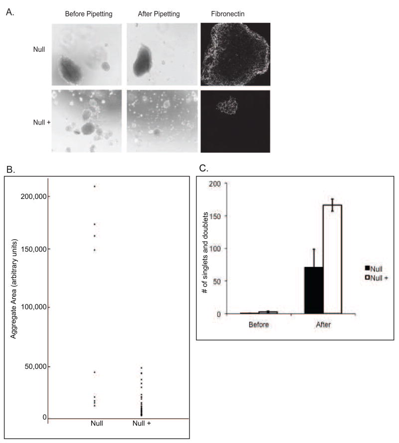
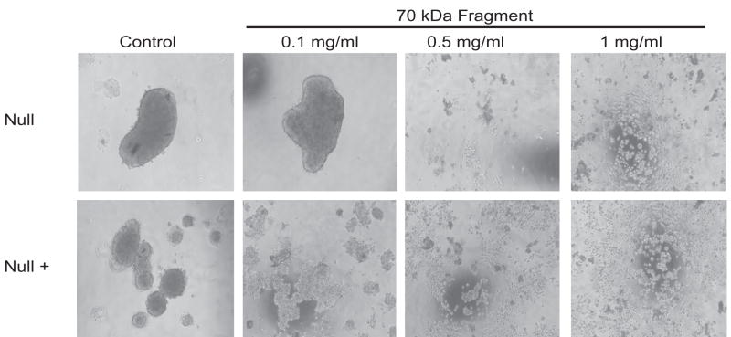
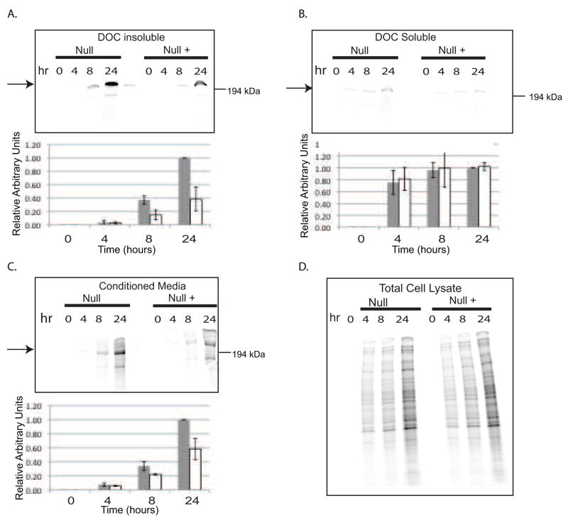
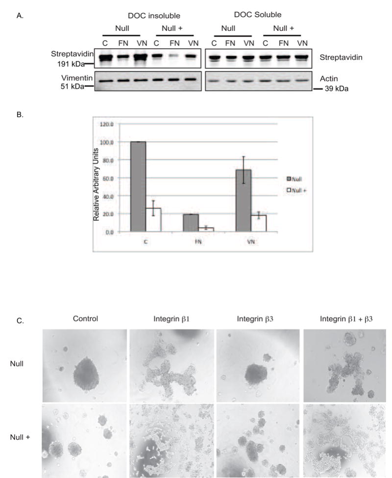
Phospholipase C-gamma1 (PLC-gamma1) mediates cell adhesion and migration through an undefined mechanism. Here, we examine the role of PLC-gamma1 in cell-matrix adhesion in a hanging drop assay of cell aggregation. Plcg1 Null (-/-) mouse embryonic fibroblasts formed aggregates that were larger and significantly more resistant to dissociation than cells in which PLC-gamma1 is re-expressed (Null+ cells). Aggregate formation could be disrupted by inhibition of fibronectin interaction with integrins, indicating that fibronectin assembly may mediate aggregate formation. Fibronectin assembly was mediated by integrin alpha5beta1 in both cell lines, while assays measuring fibronectin assembly revealed increased assembly in the Null cells. Null and Null+ cells exhibited equivalent fibronectin mRNA levels and equivalent levels of fibronectin protein in pulse-labeling experiments. However, levels of secreted fibronectin in the conditioned medium were increased in Null cells. The data implicates a negative regulatory role for PLC-gamma1 in cell aggregation by controlling the secretion of fibronectin into the media and its assembly into fibrils.
Figures

Similar articles
-
Integrin-dependent PLC-gamma1 phosphorylation mediates fibronectin-dependent adhesion.J Cell Sci. 2005 Feb 1;118(Pt 3):601-10. doi: 10.1242/jcs.01643. Epub 2005 Jan 18. J Cell Sci. 2005. PMID: 15657076
-
Alpha5beta1 integrin mediates strong tissue cohesion.J Cell Sci. 2003 Jan 15;116(Pt 2):377-86. doi: 10.1242/jcs.00231. J Cell Sci. 2003. PMID: 12482923
-
Deletion of mouse embryo fibroblast N-acetylglucosaminyltransferase V stimulates alpha5beta1 integrin expression mediated by the protein kinase C signaling pathway.J Biol Chem. 2005 Mar 4;280(9):8332-42. doi: 10.1074/jbc.M413532200. Epub 2004 Dec 22. J Biol Chem. 2005. PMID: 15615721
-
Phospholipase C-gamma1 potentiates integrin-dependent cell spreading and migration through Pyk2/paxillin activation.Cell Signal. 2007 Aug;19(8):1784-96. doi: 10.1016/j.cellsig.2007.04.002. Epub 2007 Apr 19. Cell Signal. 2007. PMID: 17531443
-
Fibronectin fibrillogenesis, a cell-mediated matrix assembly process.Matrix Biol. 2005 Sep;24(6):389-99. doi: 10.1016/j.matbio.2005.06.008. Matrix Biol. 2005. PMID: 16061370 Review.
Cited by
-
Phosphoinositide pathway and the signal transduction network in neural development.Neurosci Bull. 2012 Dec;28(6):789-800. doi: 10.1007/s12264-012-1283-x. Epub 2012 Nov 14. Neurosci Bull. 2012. PMID: 23152330 Free PMC article. Review.
-
Lypopolysaccharide downregulates the expression of selected phospholipase C genes in cultured endothelial cells.Inflammation. 2013 Aug;36(4):862-8. doi: 10.1007/s10753-013-9613-3. Inflammation. 2013. PMID: 23420070
-
Annexin A5 regulates hepatocarcinoma malignancy via CRKI/II-DOCK180-RAC1 integrin and MEK-ERK pathways.Cell Death Dis. 2018 May 25;9(6):637. doi: 10.1038/s41419-018-0685-8. Cell Death Dis. 2018. PMID: 29802377 Free PMC article.
-
Phosphorylated vimentin-triggered fibronectin matrix disaggregation enhances the dissemination of Treponema pallidum subsp. pallidum across the microvascular endothelial barrier.PLoS Pathog. 2024 Sep 3;20(9):e1012483. doi: 10.1371/journal.ppat.1012483. eCollection 2024 Sep. PLoS Pathog. 2024. PMID: 39226326 Free PMC article.
-
Regulation of retinal angiogenesis by phospholipase C-β3 signaling pathway.Exp Mol Med. 2016 Jun 17;48(6):e240. doi: 10.1038/emm.2016.39. Exp Mol Med. 2016. PMID: 27311705 Free PMC article.
References
-
- Tvorogov D, Wang XJ, Zent R, Carpenter G. Integrin-dependent PLC-gamma1 phosphorylation mediates fibronectin-dependent adhesion. Journal of cell science. 2005;118:601–610. - PubMed
-
- Kim JW, Sim SS, Kim UH, Nishibe S, Wahl MI, Carpenter G, Rhee SG. Tyrosine residues in bovine phospholipase C-gamma phosphorylated by the epidermal growth factor receptor in vitro. J Biol Chem. 1990;265:3940–3943. - PubMed
-
- Kim HK, Kim JW, Zilberstein A, Margolis B, Kim JG, Schlessinger J, Rhee SG. PDGF stimulation of inositol phospholipid hydrolysis requires PLC-gamma 1 phosphorylation on tyrosine residues 783 and 1254. Cell. 1991;65:435–441. - PubMed
Publication types
MeSH terms
Substances
Grants and funding
LinkOut - more resources
Full Text Sources
Other Literature Sources
Molecular Biology Databases
Miscellaneous
