

Cortical depth dependence and implications on the neuronal specificity of the functional apparent diffusion coefficient contrast
- PMID: 19379817
- PMCID: PMC2744342
- DOI: 10.1016/j.neuroimage.2009.04.045
Cortical depth dependence and implications on the neuronal specificity of the functional apparent diffusion coefficient contrast
Abstract
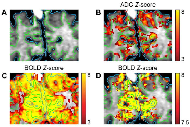
Although the blood oxygenation level-dependent (BOLD) contrast is widely used in functional MRI (fMRI), its spatial specificity is compromised by the diversity of the participating vasculature, including large draining veins. Previous studies have shown that an alternative contrast mechanism based on functional changes of the apparent diffusion coefficient (ADC) can be sensitized to small vessels more closely tied to the sites of neural activity. Such an improved functional localization, however, has not yet been demonstrated at the cortical level in humans. Here, we investigate the cortical depth dependence and neuronal specificity of the functional ADC contrast in the human primary visual cortex by performing high-resolution BOLD and ADC imaging during visual stimulation at 4 T. Our results show that, by using optimal parameters, the functional ADC changes are significantly higher in the middle cortical layers, whereas the BOLD signal changes are higher at the cortical surface and vary much less significantly across the cortex. These results are in good agreement with previous studies performed in anesthetized cats at 9.4 T and demonstrate the improved spatial specificity of the functional ADC contrast as compared to the BOLD contrast.
Figures


Similar articles
-
Sources of functional apparent diffusion coefficient changes investigated by diffusion-weighted spin-echo fMRI.Magn Reson Med. 2006 Dec;56(6):1283-92. doi: 10.1002/mrm.21074. Magn Reson Med. 2006. PMID: 17051530
-
Decreases in ADC observed in tissue areas during activation in the cat visual cortex at 9.4 T using high diffusion sensitization.Magn Reson Imaging. 2008 Sep;26(7):889-96. doi: 10.1016/j.mri.2008.01.046. Epub 2008 May 16. Magn Reson Imaging. 2008. PMID: 18486391 Free PMC article.
-
Functional changes of apparent diffusion coefficient during visual stimulation investigated by diffusion-weighted gradient-echo fMRI.Neuroimage. 2008 Jul 1;41(3):801-12. doi: 10.1016/j.neuroimage.2008.03.014. Epub 2008 Mar 20. Neuroimage. 2008. PMID: 18450483 Free PMC article.
-
Linking brain vascular physiology to hemodynamic response in ultra-high field MRI.Neuroimage. 2018 Mar;168:279-295. doi: 10.1016/j.neuroimage.2017.02.063. Epub 2017 Feb 22. Neuroimage. 2018. PMID: 28254456 Review.
-
What We Have Learned about Human Primary Visual Cortex from High Resolution Functional Magnetic Resonance Imaging.Magn Reson Med Sci. 2016;15(1):1-10. doi: 10.2463/mrms.2015-0008. Epub 2015 Jun 23. Magn Reson Med Sci. 2016. PMID: 26104083 Review.
Cited by
-
Facilitating Mitochondrial Calcium Uptake Improves Activation-Induced Cerebral Blood Flow and Behavior after mTBI.Front Syst Neurosci. 2016 Mar 8;10:19. doi: 10.3389/fnsys.2016.00019. eCollection 2016. Front Syst Neurosci. 2016. PMID: 27013987 Free PMC article.
-
Cortical layer-dependent arterial blood volume changes: improved spatial specificity relative to BOLD fMRI.Neuroimage. 2010 Jan 15;49(2):1340-9. doi: 10.1016/j.neuroimage.2009.09.061. Epub 2009 Sep 30. Neuroimage. 2010. PMID: 19800013 Free PMC article.
-
Cerebral blood volume sensitive layer-fMRI in the human auditory cortex at 7T: Challenges and capabilities.PLoS One. 2023 Feb 9;18(2):e0280855. doi: 10.1371/journal.pone.0280855. eCollection 2023. PLoS One. 2023. PMID: 36758009 Free PMC article.
References
-
- Does MD, Zhong J, Gore JC. In vivo measurement of ADC change due to intravascular susceptibility variation. Magn Reson Med. 1999;41:236–240. - PubMed
-
- Gangstead SL, Song AW. On the timing characteristics of the apparent diffusion coefficient contrast in fMRI. Magn Reson Med. 2002;48:385–388. - PubMed
-
- Jin T, Zhao F, Kim SG. Sources of functional apparent diffusion coefficient changes investigated by diffusion-weighted spin-echo fMRI. Magn Reson Med. 2006;56:1283–1292. - PubMed
-
- Lai S, Hopkins AL, Haacke EM, Li D, Wasserman BA, Buckley P, Friedman L, Meltzer H, Hedera P, Friedland R. Identification of vascular structures as a major source of signal contrast in high resolution 2D and 3D functional activation imaging of the motor cortex at 1.5T: preliminary results. Magn Reson Med. 1993;30:387–392. - PubMed
Publication types
MeSH terms
Substances
Grants and funding
LinkOut - more resources
Full Text Sources
Miscellaneous
