Review
. 2009 May;32(5):291-301.
doi: 10.1016/j.tins.2009.01.007. Epub 2009 Apr 18.

Decision by division: making cortical maps

Affiliations
Review

Decision by division: making cortical maps

Pasko Rakic et al. Trends Neurosci. 2009 May.

Abstract

In the past three decades, mounting evidence has revealed that specification of the basic cortical neuronal classes starts at the time of their final mitotic divisions in the embryonic proliferative zones. This early cell determination continues during the migration of the newborn neurons across the widening cerebral wall, and it is in the cortical plate that they attain their final positions and establish species-specific cytoarchitectonic areas. Here, the development and evolutionary expansion of the neocortex is viewed in the context of the radial unit and protomap hypotheses. A broad spectrum of findings gave insight into the pathogenesis of cortical malformations and the biological bases for the evolution of the modern human neocortex. We examine the history and evidence behind the concept of early specification of neurons and provide the latest compendium of genes and signaling molecules involved in neuronal fate determination and specification.


Figures

Figure 1
Figure 1
(A) Array of proliferative units in the VZ subjacent to the prospective primary visual cortex in the E91 monkey embryo as seen in epon-embedded tissue cut 1 μm stained with cresyl violet. Although most mitotic figures are located directly at the ventricular surface (vertical arrow), many can be found in the SVZ (horizontal double-crossed arrow). (B) Superjacent cortical plate of the same animal showing ontogenetic columns originated from the same proliferative units as illustrated in part (A). (C) Autoradiogram of an adult monkey exposed to [3H]-thymidine at E70, showing that the most intensely labeled cell (see arrow a) lies deeper in the cortex than the two progressively less labeled (arrows b and c), more superficially situated neurons, indicating inside-out sequence of their origin. (D,E) Unlabeled neurons (crossed arrows) might be interspersed among radioactive neurons (simple arrows) indicating that the ontogenetic columns originate from more than one progenitor. (Combined from Figures 1 and 2 in Ref. [22]).
Figure 2
Figure 2
3D reconstruction of migrating neurons, based originally on electron micrographs of serial sections of the monkey fetal cerebral wall modified from Ref. [4]. The more recent, animated version of this remonstration can be seen at: http://rakiclab.med.yale.edu/MigratingCorticalNeuron.html. Abbreviations: CC, corticocortical connection; CP, cortical plate; IZ, intermediate zone; MA, monoamine; MN, migrating neuron; MZ, marginal zone; NB, nucleus basalis; RG, radial glia; SP, subplate; TR, thalamic radiation; VZ, ventricular zone.
Figure 3
Figure 3
Patterning centers around the neocortex are responsible for establishing morphogenetic gradients that govern areal identity in the cortex. The table lists key molecules and their corresponding expression pattern. An extensive list is available at: http://rakiclab.med.yale.edu/pages/molecules.php. Abbreviations: Foxg1, forkhead box G1 (also known as brain factor 1 [BF1]); Nrg1, Nrg2 and Nrg3, neuregulin 1, 2 and 3; Otx2, orthodenticle homeobox 2; Spry1, sprouty homolog 1; TGFα, transforming growth factor α.
Figure 4
Figure 4
Graded expression patterns of several factors thought to be responsible for shaping the ultimate neocortical landscape that is highly partitioned and specialized. A greater list is available at: http://rakiclab.med.yale.edu/pages/molecules.php. Abbreviations: Id2, Inhibitor of DNA binding 2; Lhx2, LIM homeobox 2; Lmo3, LIM domain only 3; ROR-β retinoid-related orphan receptor β.
Figure 5
Figure 5
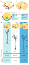
Re-utilization of the protomap in the postnatal brain and glia. (a) Example of molecules involved in specification of the embryonic forebrain. (b) These molecules are similarly utilized in the postnatal forebrain for the generation of olfactory bulb (OB) neurons and glia in the OB and other forebrain regions as shown by several groups. (c) Summary of results from Ref. [126] showing that the combinatorial progenitor code (in this case, combinations of Pax6, Nkx6.1 and Nkx2.2 transcription factor expression) in the embryonic spinal cord pre-specify the eventual areal (VA1,VA2 or VA3) and secretion (reelin, Slit or both) subtype of astrocytes into three subclasses. (d) The location of generation of OB granule cells correlates with the location of the progenitor cells in the postnatal subependymal (SEZ) zone. Neonatal granule cells born in the anterior SEZ (aSEZ) typically have dendrites that branch in the superficial external plexiform layer (EPL), whereas posterior (SEZ)-derived granule cells branch in the deep EPL. Postnatally born aSEZ-derived granule cells preferentially branch in the EPL. See text for discussion of these results from Lois and colleagues [127]. Abbreviations: Ctx, cortex; Dbx1, developing brain homeobox 1; Emx1, empty spiracles homeobox 1; GCL, granule cell layer; GL, glomerular layer; Gsh2, genomic screen homeobox 2; LGE, lateral ganglionic eminence; MCL, mitral cell layer; MGE, medial ganglionic eminence; Nkx2.2, NK2 homeobox 1; Nkx6.1, NK6 homeobox 1; pA1, pA2 and pA3, progenitor domain astrocyte subtypes A1, A2 and A3; Pax6, paired box 6; pSEZ, posterior subependymal zone; RMS, rostral migratory stream; Str, striatum.

References

    1. Kuan CY, et al. Mechanisms of programmed cell death in the developing brain. Trends Neurosci. 2000;23:291–297. - PubMed
    1. Rakic P. Neuronal–glial interaction during brain development. Trends Neurosci. 1981;4:184–187.
    1. Rakic P. Mechanism of ocular dominance segregation in the lateral geniculate nucleus: competitive elimination hypothesis. Trends Neurosci. 1986;9:11–15.
    1. Rakic P. A small step for the cell, a giant leap for mankind: a hypothesis of neocortical expansion during evolution. Trends Neurosci. 1995;18:383–388. - PubMed
    1. Sarkisian MR, et al. Trouble making the first move: interpreting arrested neuronal migration in the cerebral cortex. Trends Neurosci. 2008;31:54–61. - PubMed

Publication types