Review
. 2009 May;50 Suppl 1(Suppl 1):106S-21S.
doi: 10.2967/jnumed.108.057281. Epub 2009 Apr 20.

Radiopharmaceuticals in preclinical and clinical development for monitoring of therapy with PET

Affiliations
Review

Radiopharmaceuticals in preclinical and clinical development for monitoring of therapy with PET

Mark P S Dunphy et al. J Nucl Med. 2009 May.

Abstract

This review article discusses PET agents, other than (18)F-FDG, with the potential to monitor the response to therapy before, during, or after therapeutic intervention. This review deals primarily with non-(18)F-FDG PET tracers that are in the final stages of preclinical development or in the early stages of clinical application for monitoring the therapeutic response. Four sections related to the nature of the tracers are included: radiotracers of DNA synthesis, such as the 2 most promising agents, the thymidine analogs 3'-(18)F-fluoro-3'-deoxythymidine and (18)F-1-(2'-deoxy-2'-fluoro-beta-d-arabinofuranosyl)thymine; agents for PET imaging of hypoxia within tumors, such as (60/62/64)Cu-labeled diacetyl-bis(N(4)-methylthiosemicarbazone) and (18)F-fluoromisonidazole; amino acids for PET imaging, including the most popular such agent, l-[methyl-(11)C]methionine; and agents for the imaging of tumor expression of androgen and estrogen receptors, such as 16beta-(18)F-fluoro-5alpha-dihydrotestosterone and 16alpha-(18)F-fluoro-17beta-estradiol, respectively.


Figures

FIGURE 1
FIGURE 1
(A) 18F-FLT. (B) 18F-FMAU.
FIGURE 2
FIGURE 2
Newly diagnosed glioblastoma. (A) MRI (contrast-enhanced T1-weighted image) shows large area of contrast enhancement in right frontal lobe. (B and C) Both 18F-FDG PET (B) and 18F-FLT PET (C) show increased uptake in same area. (Reprinted with permission of (189).)
FIGURE 3
FIGURE 3
(A) 64Cu-ATSM. (B) 18F-FMISO.
FIGURE 4
FIGURE 4
Progression-free survival (left) and cause-specific survival (right) determined from 60Cu-ATSM uptake by Kaplan–Meier method. ○ = event in patients with T/M of ≤3.5; ∇ = event in patients with T/M of >3.5. (Reprinted with permission of (83).)
FIGURE 5
FIGURE 5
(A) Transaxial CT (top left) and 18F-FDG PET (top right) images of pelvis show intense 18F-FDG uptake within known cervical tumor at site of cervical mass seen on CT. Transaxial 30- to 60-min summed images of 60Cu-ATSM PET (bottom left) and 64Cu-ATSM PET (bottom right) of pelvis at same level demonstrate mildly increased uptake within known primary cervical tumor. There are similar patterns of 60Cu-ATSM and 64Cu-ATSM uptake within tumor. (B) Transaxial coregistered 18F-FDG PET/CT (top left) and 18F-FDG PET (top right) images of pelvis show intense 18F-FDG uptake within known cervical tumor at site of cervical mass seen on CT. Transaxial 30- to 60-min summed images of 60Cu-ATSM PET (bottom left) and 64Cu-ATSM PET (bottom right) of pelvis at same level demonstrate markedly increased uptake within known primary cervical tumor. There are similar patterns of 60Cu-ATSM and 64Cu-ATSM uptake within tumor. (Reprinted with permission of (86).)
FIGURE 6
FIGURE 6
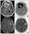
(A and B) Bifrontal glioblastoma multiforme imaged after biopsy in 55-y-old woman. (A) MRI (gadolinium-enhanced T1-weighted image [T1Gd]) shows large, contrast-enhancing, irregular ring-shaped tumor with necrotic center. Non–contrast-enhanced volume was 20 cm3, T1Gd volume was 80 cm3, and T2-weighted volume was 167 cm3. (B) 18F-FMISO image through same plane. HV was 129 cm3, and T/Bmax ratio was 3.0. (C and D) Left temporal glioblastoma multiforme imaged after gross total resection in 53-y-old man. (C) MRI (T1Gd) shows only blood products and no residual contrast-positive disease. Non–contrast-enhanced volume was 1 cm3, T1Gd volume was 7 cm3, and T2-weighted volume was 37 cm3. (D) 18F-FMISO image through same plane. HV was 5.3 cm3, and T/Bmax ratio was 1.6. (Reprinted with permission of (97).)
FIGURE 7
FIGURE 7
Imaging of 49-y-old woman who had been previously treated for glioblastoma multiforme with tumor resection and conventional radiotherapy at dose of 60 Gy. (A) T1-weighted MR image obtained with contrast medium 13 mo after initial surgery showed contrast-enhanced lesion in left frontal lobe. (B) 11C-MET PET image showed obvious accumulation of tracer corresponding to abnormality on MR image. Mean lesion-to-normal tissue ratio was 1.70. Recurrent tumor was pathologically confirmed by second surgery. (Reprinted with permission of (106).)
FIGURE 8
FIGURE 8
(A) 18F-FES. (B) 18F-FDHT.
FIGURE 9
FIGURE 9
Baseline tumor 18F-FES (left) and percentage change in tumor 18F-FDG (right) uptake after estradiol challenge in patients who responded and patients who did not respond to endocrine therapy. (Reprinted with permission of (140).)
FIGURE 10
FIGURE 10
18F-FDHT (A) and 18F-FDG (B) scans displayed in maximum-intensity-projection format. This figure demonstrates the contrasting metabolism of the 2 tracers in a patient with metastatic prostate cancer. (Reprinted with permission of (158).)

References

    1. Seidlin S, Marinelli L, Oshry E. Radioactive iodine therapy: effect on functioning metastases of adenocarcinoma of the thyroid. JAMA. 1946;132:838–847. - PubMed
    1. Dunn JT, Dunn AD. Update on intrathyroidal iodine metabolism. Thyroid. 2001;11:407–414. - PubMed
    1. Robbins RJ, Wan Q, Grewal RK, et al. Real-time prognosis for metastatic thyroid carcinoma based on 2-[18F]fluoro-2-deoxy-d-glucose-positron emission tomography scanning. J Clin Endocrinol Metab. 2006;91:498–505. - PubMed
    1. Hadj-Djilani NL, Lebtahi NE, Delaloye AB, Laurini R, Beck D. Diagnosis and follow-up of neuroblastoma by means of iodine-123 metaiodobenzylguanidine scintigraphy and bone scan, and the influence of histology. Eur J Nucl Med. 1995;22:322–329. - PubMed
    1. Lastoria S, Maurea S, Caraco C, et al. Iodine-131 metaiodobenzylguanidine scintigraphy for localization of lesions in children with neuroblastoma: comparison with computed tomography and ultrasonography. Eur J Nucl Med. 1993;20:1161–1167. - PubMed

Publication types

LinkOut - more resources