. 2009 Jun 26;284(26):17465-74.
doi: 10.1074/jbc.M109.007179. Epub 2009 Apr 20.

Solution structures of cytosolic RNA sensor MDA5 and LGP2 C-terminal domains: identification of the RNA recognition loop in RIG-I-like receptors

Affiliations

Solution structures of cytosolic RNA sensor MDA5 and LGP2 C-terminal domains: identification of the RNA recognition loop in RIG-I-like receptors

Kiyohiro Takahasi et al. J Biol Chem. .

Abstract

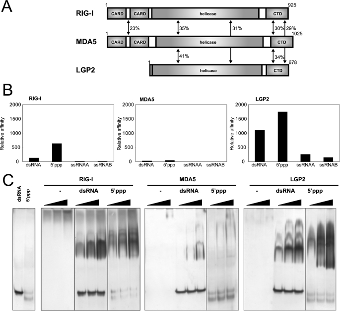
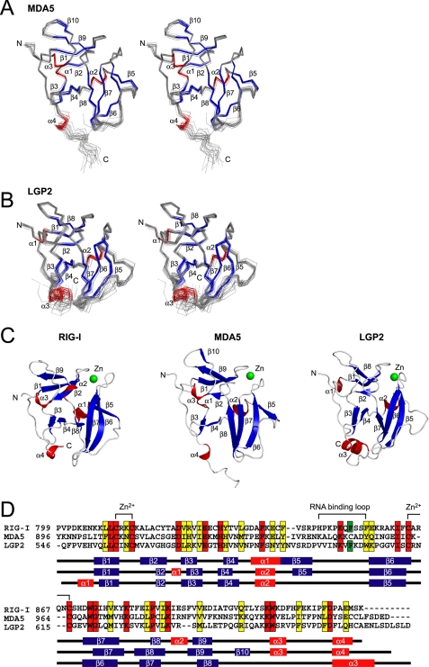
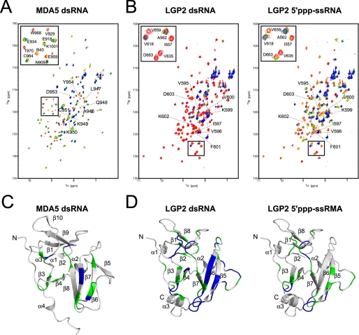
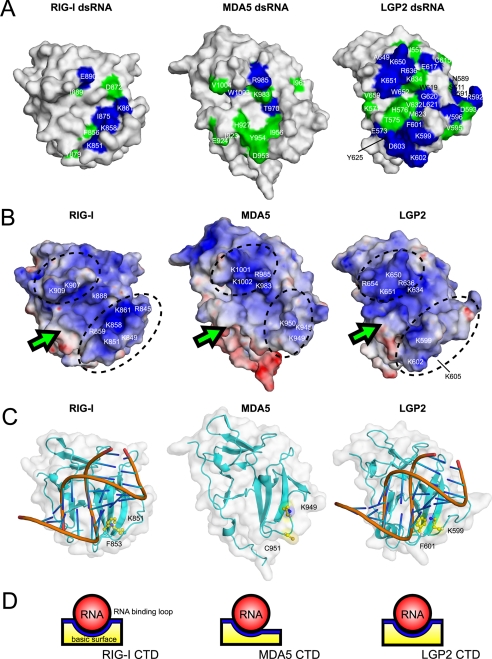
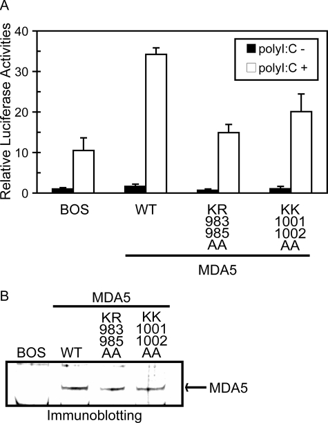
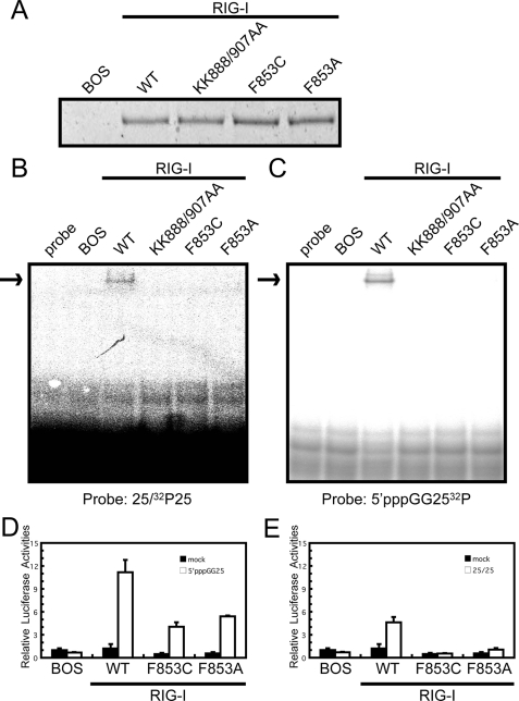
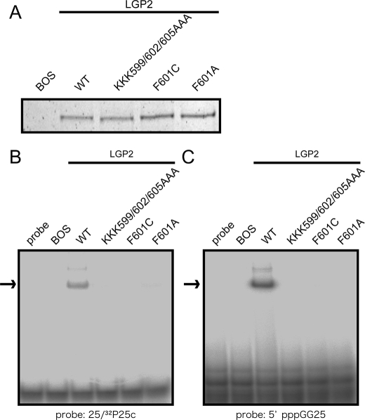
The RIG-I like receptor (RLR) comprises three homologues: RIG-I (retinoic acid-inducible gene I), MDA5 (melanoma differentiation-associated gene 5), and LGP2 (laboratory of genetics and physiology 2). Each RLR senses different viral infections by recognizing replicating viral RNA in the cytoplasm. The RLR contains a conserved C-terminal domain (CTD), which is responsible for the binding specificity to the viral RNAs, including double-stranded RNA (dsRNA) and 5'-triphosphated single-stranded RNA (5'ppp-ssRNA). Here, the solution structures of the MDA5 and LGP2 CTD domains were solved by NMR and compared with those of RIG-I CTD. The CTD domains each have a similar fold and a similar basic surface but there is the distinct structural feature of a RNA binding loop; The LGP2 and RIG-I CTD domains have a large basic surface, one bank of which is formed by the RNA binding loop. MDA5 also has a large basic surface that is extensively flat due to open conformation of the RNA binding loop. The NMR chemical shift perturbation study showed that dsRNA and 5'ppp-ssRNA are bound to the basic surface of LGP2 CTD, whereas dsRNA is bound to the basic surface of MDA5 CTD but much more weakly, indicating that the conformation of the RNA binding loop is responsible for the sensitivity to dsRNA and 5'ppp-ssRNA. Mutation study of the basic surface and the RNA binding loop supports the conclusion from the structure studies. Thus, the CTD is responsible for the binding affinity to the viral RNAs.


Figures

FIGURE 1.
FIGURE 1.
Functional analysis of RLR CTDs. A, domain structures of RIG-I, MDA5, and LGP2. Sequence identities of each domain among the RLRs are indicated. B, RNA-binding activity of RLR CTDs determined by SPR. GST-RIG-I CTD, GST-MDA5 CTD, and GST-LGP2 CTD were captured by the anti-GST anti-body immobilized onto the sensor chip, then dsRNA, 5′ppp-ssRNA, and two ssRNAs (ssRNAA and ssRNAB) were injected. Each resonance unit of RNA bound to a GST fused protein are standardized by molecular weight of the RNAs, then normalized by the resonance unit and molecular weight of the captured GST-fused protein. Normalized data are summarized and shown as bar graphs. RNAs bound to RIG-I, MDA5, and LGP2 are indicated from left to right in the panels. C, RNA-binding activity of RLR CTDs determined by EMSA. CTDs without the GST tag were prepared and subjected to EMSA. Increasing amounts of CTD (10, 20, and 40 pm) were reacted with the indicated probe and analyzed by native PAGE. The gels were silver-stained to visualize protein and RNA probe. −, no RNA; dsRNA, 25/25c probe; 5ppp: 5′pppGG25 probe.
FIGURE 2.
FIGURE 2.
Solution structure of MDA5 CTD and LGP2 CTD. A and B, best fit superposition of the backbone atoms of 20 NMR-derived MDA5 CTD (A) and LGP2 CTD (B). Structures are shown in stereo. β-Strands and α-helices are shown in blue and red, respectively. C, ribbon diagrams of the structure of RIG-I CTD, MDA5 CTD, and LGP2 CTD (left to right). Secondary structure elements are labeled. The figure was prepared using PyMOL. D, sequence alignment of human RIG-I, MDA5, and LGP2 CTDs. ClustalX was used to align the sequences. The secondary structure elements of each CTD are indicated below the alignment. The amino acids in red and yellow indicate conserved (red) and type-conserved (yellow) residues with the Zn2+ binding Cys-X-X-Cys motifs and RNA binding loop. The Phe residues conserved in RIG-I and LGP2 in the RNA binding loop are colored green.
FIGURE 3.
FIGURE 3.
NMR titration results for MDA5 CTD and LGP2 CTD. A, NMR titration of MDA5 CTD with dsRNA. An overlay of 1H-15N HSQC spectra are shown in red, yellow, green, and blue at 0, 0.25, 0.5, and 1.0 equivalents (molar ratio of dsRNA to proteins), respectively, where the residues in the RNA binding loop are labeled. The inset shows the excerpt of the enclosed region of the spectrum with assignment. B, NMR titration of LGP2 CTD with dsRNA (left panel) and 5′ppp-ssRNA (right panel). The figures are prepared in the same manner as in A. C, mapping of the residues of MDA5 CTD affected by the addition of dsRNA on the ribbon diagram. The residues whose peaks disappeared on addition of 0.25 and 0.5 equivalent molar ratios of dsRNA to CTD are colored blue and green, respectively. D, mapping of the residues of LGP2 CTD affected by the addition of dsRNA and 5′ppp-ssRNA (left and right panels) on the ribbon diagram, colors are the same as in C. The orientations are the same as in Fig. 2, A and B.
FIGURE 4.
FIGURE 4.
Structural comparisons of RLR CTDs. A, surface representation of the dsRNA affected surface of RLR CTDs (left to right, RIG-I CTD, MDA5 CTD, and LGP2 CTD). The residues that disappeared in NMR titration experiments upon addition of 0.25 and 0.5 equivalents of dsRNA are colored in blue and green, respectively. The residue numbers are also shown. The data of the dsRNA affected surface of RIG-I CTD was derived from our previous study (8). B, electrostatic surface potentials of the RLR CTDs (left to right, RIG-I CTD, MDA5 CTD, and LGP2 CTD). Dotted circles indicate the banks surrounding the basic surface. C, left, dsRNA-bound model of RIG-I CTD. Right, dsRNA-bound model of LGP2 CTD. The Lys and Phe residues in the RNA binding loop conserved in RIG-I and LGP2 are shown in ball-and-stick models and labeled. Middle, structure of MDA5 CTD, the residues corresponding to the Lys-851 and Phe-853 in RIG-I are shown. All figures are shown in the same structural orientation as in Fig. 2A. D, schematic diagrams of dsRNA bound to the basic surface of RIG-I CTD (left), MDA5 CTD (mid), and LGP2 CTD (right) viewed from the arrows indicated in Fig. 4B.
FIGURE 5.
FIGURE 5.
Functional analysis of basic residue mutations of MDA5 on the basic surface. A, MDA5−/− mouse embryo fibroblasts were transfected with the reporter gene, p-125Luc, and pRL-tk, together with the expression vector for MDA5 and mutants. Cells were stimulated by transfection with poly(I:C) and subjected to a dual-luciferase assay. The values are the means ± S.D. from triplicate experiments. The relative luciferase activity was calculated by considering the luciferase activity from cells transfected with empty vector (BOS) as 1.0. The cell lysates were analyzed for expression of MDA5 and mutants by Immunoblotting (B).
FIGURE 6.
FIGURE 6.
Functional analysis of the basic surface and the RNA binding loop mutants of RIG-I. Full-length wild type RIG-I and mutants were produced in 293T cells and purified by anti FLAG antibody. A, silver staining of the purified recombinant RIG-I. The recombinant RIG-I was subjected to EMSA using 32P-labeled dsRNA (B; 25/25c) or 5′ppp-ssRNA (C; 5′pppGG25) as probe. D and E, interferon-β promoter activation by wt RIG-I and mutants. RIG-I−/− mouse embryo fibroblasts were transfected with the reporter gene, p-125Luc, and pRL-tk, together with the expression vector for RIG-I and mutants. Cells were stimulated by transfection with 25/25c (D) or 5′pppGG25 (E) and subjected to a dual-luciferase assay. The values are the means ± S.D. from triplicate experiments. The relative luciferase activity was calculated by considering the luciferase activity from cells transfected with empty vector (BOS) as 1.0.
FIGURE 7.
FIGURE 7.
Effect of the mutations of LGP2 on RNA recognition. Full-length wild-type LGP2 and mutants on the RNA binding loop were produced in 293T cells and purified by anti FLAG antibody. A, silver staining of the purified recombinant LGP2. The recombinant LGP2 were subjected to EMSA using (B) 32P-labeled dsRNA (25/25c) or (C) 5′ppp-ssRNA (C; 5′pppGG25) as probe. The arrows indicate the RNA-LGP2 complex.

Similar articles

Cited by

References

    1. Akira S., Uematsu S., Takeuchi O. ( 2006) Cell 124, 783– 801 - PubMed
    1. Yoneyama M., Kikuchi M., Natsukawa T., Shinobu N., Imaizumi T., Miyagishi M., Taira K., Akira S., Fujita T. ( 2004) Nat. Immunol. 5, 730– 737 - PubMed
    1. Hornung V., Ellegast J., Kim S., Brzózka K., Jung A., Kato H., Poeck H., Akira S., Conzelmann K. K., Schlee M., Endres S., Hartmann G. ( 2006) Science 314, 994– 997 - PubMed
    1. Pichlmair A., Schulz O., Tan C. P., Näslund T. I., Liljeström P., Weber F., Reis E., Sousa C. ( 2006) Science 314, 997– 1001 - PubMed
    1. Kato H., Takeuchi O., Sato S., Yoneyama M., Yamamoto M., Matsui K., Uematsu S., Jung A., Kawai T., Ishii K. J., Yamaguchi O., Otsu K., Tsujimura T., Koh C. S., Reis e Sousa C., Matsuura Y., Fujita T., Akira S. ( 2006) Nature 441, 101– 105 - PubMed

Publication types

MeSH terms