. 2009 Jun 5;284(23):15894-902.
doi: 10.1074/jbc.M109.009589. Epub 2009 Apr 20.

Membrane repair defects in muscular dystrophy are linked to altered interaction between MG53, caveolin-3, and dysferlin

Affiliations

Membrane repair defects in muscular dystrophy are linked to altered interaction between MG53, caveolin-3, and dysferlin

Chuanxi Cai et al. J Biol Chem. .

Abstract

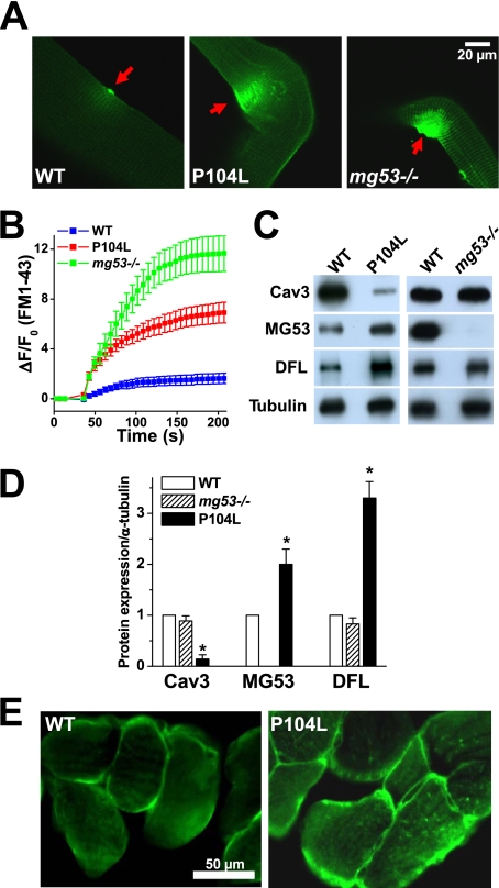
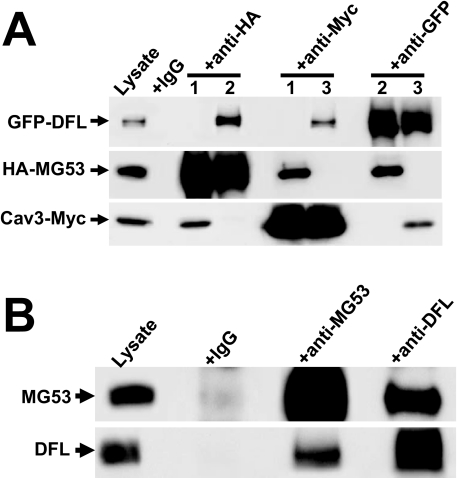
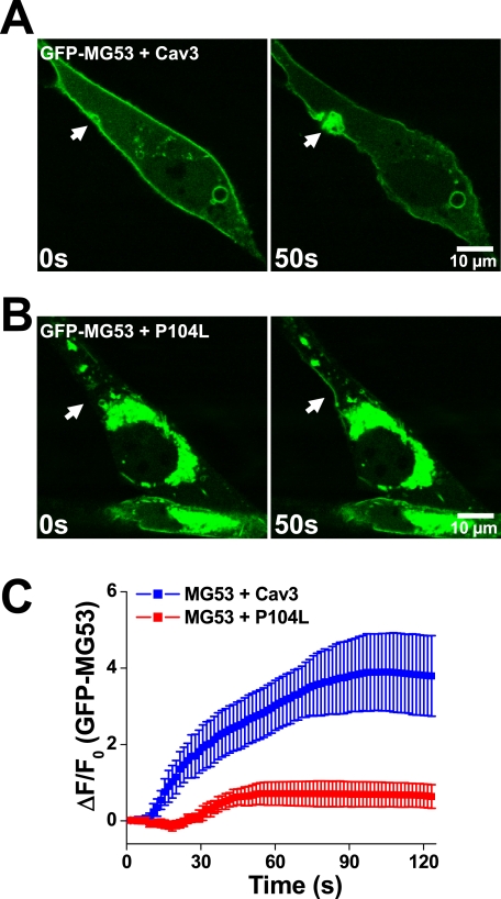
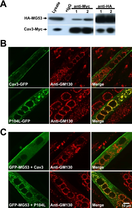
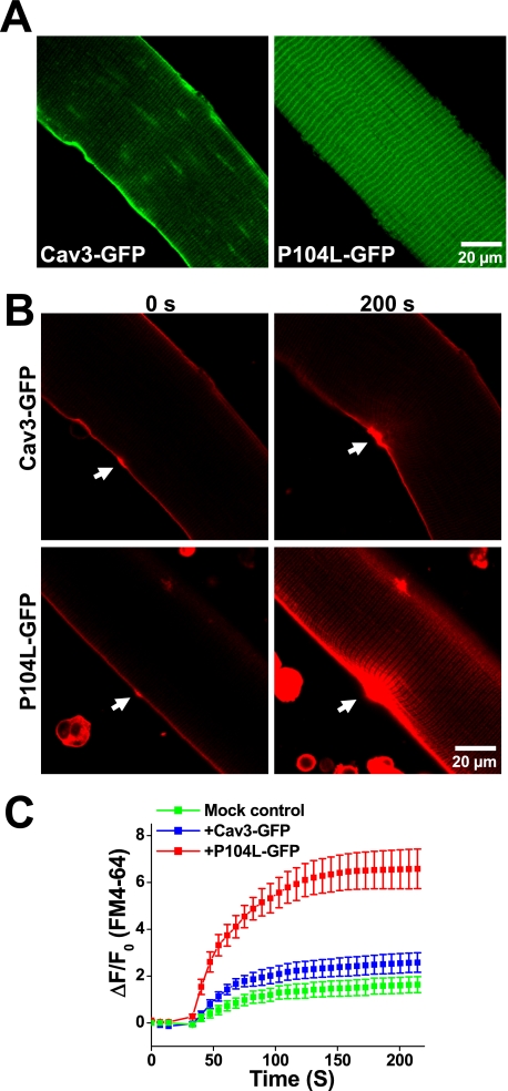
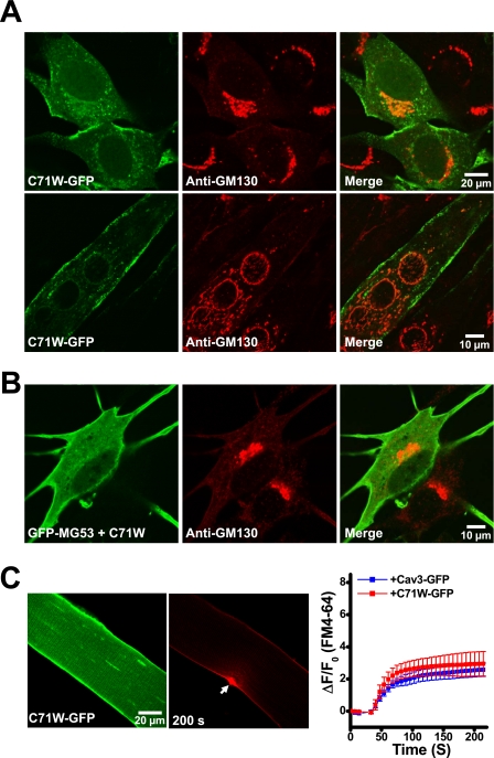
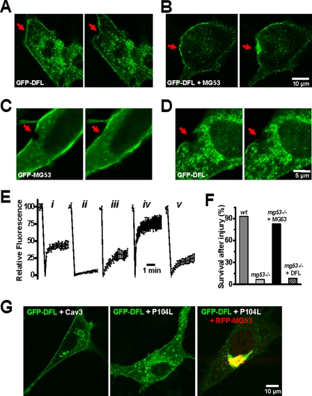
Defective membrane repair can contribute to the progression of muscular dystrophy. Although mutations in caveolin-3 (Cav3) and dysferlin are linked to muscular dystrophy in human patients, the molecular mechanism underlying the functional interplay between Cav3 and dysferlin in membrane repair of muscle physiology and disease has not been fully resolved. We recently discovered that mitsugumin 53 (MG53), a muscle-specific TRIM (Tri-partite motif) family protein (TRIM72), contributes to intracellular vesicle trafficking and is an essential component of the membrane repair machinery in striated muscle. Here we show that MG53 interacts with dysferlin and Cav3 to regulate membrane repair in skeletal muscle. MG53 mediates active trafficking of intracellular vesicles to the sarcolemma and is required for movement of dysferlin to sites of cell injury during repair patch formation. Mutations in Cav3 (P104L, R26Q) that cause retention of Cav3 in Golgi apparatus result in aberrant localization of MG53 and dysferlin in a dominant-negative fashion, leading to defective membrane repair. Our data reveal that a molecular complex formed by MG53, dysferlin, and Cav3 is essential for repair of muscle membrane damage and also provide a therapeutic target for treatment of muscular and cardiovascular diseases that are linked to compromised membrane repair.


Figures

FIGURE 1.
FIGURE 1.
Membrane repair defects and altered localization of MG53 in P104L-Cav3 skeletal muscle. A, entry of FM1-43 fluorescent dye into isolated FDB muscle fibers from WT (left), P104L transgenic (middle), or mg53−/− mice (right) following UV laser-induced damage of the sarcolemmal membrane. B, summary time course data of accumulation of FM1-43 dye in UV-damaged FDB muscle fibers derived from WT (n = 30), P104L transgenic (n = 21), or mg53−/− (n = 18) muscle fibers. Data are listed as mean ± S.E. (error bars). C, Western blot shows expression level of Cav3, MG53, and dysferlin (DFL) in WT, P104L transgenic, and mg53−/− gastrocnemius muscle. Consistent with previous observations, expression of Cav3 is reduced in the P104L transgenic muscle (22). 10 μg/lane of total protein was loaded. α-Tubulin was used as a loading control. D, quantitative analysis of protein expression level relative to α-tubulin for Cav3, MG53, and dysferlin. The expression of Cav3, MG53, and dysferlin in wild-type muscle was set as 1, and the level of α-tubulin was used as a loading control. Data are mean ± S.E. (error bars) (n = 3 independent experiments). *, p < 0.01. E, immunostaining of cross sections from WT and P104L transgenic mouse skeletal muscle using polyclonal anti-MG53 antibody shows altered localization of MG53 in P104L muscle.
FIGURE 2.
FIGURE 2.
Co-IP reveals physical interaction between MG53, Cav3, and DFL. A, C2C12 myoblast cells were co-expressed with HA-MG53 and Cav3-Myc (lanes labeled 1), HA-MG53 with GFP-DFL (lanes labeled 2), or Cav3-Myc with GFP-DFL (lanes labeled 3). 24 h after transfection, cell lysates were immunoprecipitated and blotted with antibody to anti-GFP, anti-HA, or anti-Myc. Co-IP of DFL and Cav3 was detected with anti-HA; Co-IP of MG53 and DFL was detected with anti-Myc; and Co-IP of MG53 and Cav3 was detected with anti-GFP. Cell lysates incubated with normal mouse IgG served as control. B, physical interaction between MG53 and DFL is also observed in gastrocnemius muscle from the WT mice. Co-IP of DFL is detected with anti-MG53, and Co-IP of MG53 is detected with anti-DFL. Preimmune IgG was used as a negative control.
FIGURE 3.
FIGURE 3.
Defective movement of GFP-MG53-containing vesicles to acute membrane injury sites with co-expression of P104L-Cav3. A, C2C12 myoblast cells transfected with GFP-MG53 and Cav3 were subjected to penetration by a microelectrode. Rapid recruitment of GFP-MG5-containing vesicles toward the injury site (arrow) was observed (see supplemental Movie 1). B, co-transfection of P104L-Cav3 and GFP-MG53 in C2C12 myoblast cells leads to mistargeting of GFP-MG53 in intracellular compartments and compromised GFP-MG53 translocation to injury sites (see supplemental Movie 2). C, summary data for time-dependent accumulation of GFP-MG53 at the injury sites were plotted for C2C12 cells co-transfected with GFP-MG53 and Cav3 or GFP-MG53 and P104L-Cav2. Data are mean ± S.E. (error bars) for n = 18 cells.
FIGURE 4.
FIGURE 4.
P104L-Cav3 causes retention of MG53 at the Golgi network. A, C2C12 cells were co-transfected with HA-MG53 and either Cav3-Myc (lane 1) or P104L-Myc (lane 2). 24 h after transfection, cell lysates were immunoprecipitated and Western blotted with anti-HA or anti-Myc antibody. Cell lysates incubated with normal mouse IgG served as control. B, C2C12 myotubes transfected with Cav3-GFP or P104L-GFP were subjected to immunostaining with anti-GM130, a molecular marker for Golgi apparatus. Confocal images show that Cav3-GFP displays a plasma membrane pattern that does not overlap with GM130 staining (upper). P104L-GFP displays an intracellular localization pattern that co-localizes with GM130 (lower). C, C2C12 myotubes co-transfected with GFP-MG53 and Cav3-Myc or P104L-Myc were subjected to immunostaining with anti-GM130. Confocal images show that co-expression of Cav3 with GFP-MG53 does not impact the plasma membrane-tethering pattern of MG53 (upper), whereas co-expression of P104L with GFP-MG53 results in retention of MG53 at the Golgi apparatus (lower). These are representative images from >30 different cells.
FIGURE 5.
FIGURE 5.
Dominant effect of P104L-Cav3 produces defective membrane repair in native skeletal muscle. A, FDB muscle fibers from WT mice were transfected by in vivo electroporation to allow for transient expression of Cav3-GFP (left) and P104L-GFP (right) that were visualized by confocal microscopy. Confocal images showed that Cav3-GFP mainly targets to the sarcolemmal membrane, whereas P104L-GFP displayed a pattern indicative of intracellular retention of the mutant Cav3 protein. B, measurement of FM4-64 entry revealed severe defects in membrane repair capacity in fibers expressing P104L-GFP (lower), where excessive FM4-64 dye entry is observed following UV laser wounding (arrows) when compared with Cav3-GFP-expressing fibers (upper). C, quantitative assay of FM4-64 dye entry into skeletal muscle transiently expressing Cav3-GFP (blue trace) or P104L-GFP (red trace) or untransfected fibers on the same dish (Mock control, green trace). Data are means ± S.E. (error bars) for n = 18 fibers for each group from 3 independent electroporation with WT mice.
FIGURE 6.
FIGURE 6.
C71W-Cav3 does not affect membrane repair capacity or MG53 localization in skeletal muscle. A, C2C12 myoblast or myotube cells transfected with C71W-GFP were subjected to immunostaining with anti-GM130. Confocal images show that C71W-GFP does not co-localize with GM130 in C2C12 cells at either the myoblast stage (upper) or the differentiated myotubes (lower). B, C2C12 myoblast cells co-transfected with GFP-MG53 and C71W-Myc were subjected to immunostaining with anti-GM130. Confocal images show that co-expression of C71W-Cav3 with GFP-MG53 does not cause retention of GFP-MG53 in the Golgi apparatus. C, FDB muscle fibers from WT mice were transfected by in vivo electroporation to allow for transient expression of C71W-GFP (left). Confocal images show that C71W-GFP mainly target to the sarcolemmal membrane. Measurement of FM4-64 entry (middle) shows that transient expression of C71W-GFP in adult WT skeletal muscle produced a similar degree of FM4-64 dye entry following UV laser wounding (arrows) as Cav3-GFP-expressing fibers (right panel, n = 18). Error bars indicate S.E.
FIGURE 7.
FIGURE 7.
MG53 is required for dysferlin function in membrane repair. A, acute microelectrode penetration into the plasma membrane did not cause redistribution of GFP-dysferlin toward the injury site in C2C12 myoblasts (n = 31). B, co-expression of GFP-dysferlin and MG53 can facilitate the recruitment of GFP-dysferlin toward the injury site in C2C12 myoblasts (n = 29). Right panels in A and B are 60 s after acute mechanic injury. For details of the dynamic membrane repair process, see supplemental Movie 3 and supplemental Movie 4. C, laser-induced bleaching of GFP fluorescence at the C2C12 plasma membrane is accompanied with rapid recovery of GFP-MG53 toward the damage site (arrow, n = 20). D, C2C12 myoblasts expressing GFP-dysferlin do not display recovery following UV bleaching (n = 20). Right panels in C and D are 100 s after laser bleaching. E, summary of fluorescence recovery following photo-bleaching in C2C12 myoblasts expressing GFP-MG53 (n = 20) (panel i); C2C12 myoblasts expressing GFP-dysferlin (n = 20) (panel ii); C2C12 myoblasts co-expressing GFP-dysferlin and MG53 (n = 45) (panel iii); C2C12 myotubes expressing GFP-MG53 at 7 days of differentiation (n = 35) (panel iv); and C2C12 myotubes expressing GFP-dysferlin at 7 days of differentiation (n = 35) (panel v). Error bars indicate S.E. F, primary cultured WT or mg53−/− myotubes transfected with either GFP-MG53 or GFP-DFL were subjected to mechanical injury by microelectrode penetration. Survival of mg53−/− myotubes was greatly compromised (n = 3/38) when compared with WT myotubes (n = 32/35) due to the excessive entry of extracellular Ca2+-inducing myotube contraction. Expression of MG53 rescues mg53−/− myotube survival (n = 26/32), whereas DFL does not (n = 3/36). G, mg53−/− myoblast cells were transfected with GFP-DFL and Cav3 (left), GFP-DFL and P104L-Cav3 (middle), or GFP-DFL, RFP-MG53, and P104L-Cav3 (right). These confocal images were representative of n > 20 cells examined.

References

    1. McNeil P. L., Kirchhausen T. ( 2005) Nat. Rev. Mol. Cell Biol. 6, 499– 505 - PubMed
    1. Towler M. C., Kaufman S. J., Brodsky F. M. ( 2004) Traffic 5, 129– 139 - PubMed
    1. Glover L., Brown R. H., Jr.( 2007) Traffic 8, 785– 794 - PubMed
    1. Steinhardt R. A., Bi G., Alderton J. M. ( 1994) Science 263, 390– 393 - PubMed
    1. Miyake K., McNeil P. L. ( 1995) J. Cell Biol. 131, 1737– 1745 - PMC - PubMed

Publication types

MeSH terms