. 2009 May 11;206(5):991-9.
doi: 10.1084/jem.20090303. Epub 2009 Apr 20.

T follicular helper cells differentiate from Th2 cells in response to helminth antigens

Affiliations

T follicular helper cells differentiate from Th2 cells in response to helminth antigens

Arielle Glatman Zaretsky et al. J Exp Med. .

Abstract

The relationship of T follicular helper (TFH) cells to other T helper (Th) subsets is controversial. We find that after helminth infection, or immunization with helminth antigens, reactive lymphoid organs of 4get IL-4/GFP reporter mice contain populations of IL-4/GFP-expressing CD4(+) T cells that display the TFH markers CXCR5, PD-1, and ICOS. These TFH cells express the canonical TFH markers BCL6 and IL-21, but also GATA3, the master regulator of Th2 cell differentiation. Consistent with a relationship between Th2 and TFH cells, IL-4 protein production, reported by expression of huCD2 in IL-4 dual reporter (4get/KN2) mice, was a robust marker of TFH cells in LNs responding to helminth antigens. Moreover, the majority of huCD2/IL-4-producing Th cells were found within B cell follicles, consistent with their definition as TFH cells. TFH cell development after immunization failed to occur in mice lacking B cells or CD154. The relationship of TFH cells to the Th2 lineage was confirmed when TFH cells were found to develop from CXCR5(-) PD-1(-) IL-4/GFP(+) CD4(+) T cells after their transfer into naive mice and antigen challenge in vivo.


Figures
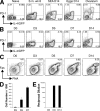
Figure 1.
Figure 1.
PD-1+ Th2 cells and germinal center B cells develop in response to helminth infections or immunization with Th2 response inducing antigens/adjuvants. (A) 4get mice were infected with S. mansoni or immunized s.c. with the antigens indicated. Various times thereafter (see Results and Discussion), cells from reactive LNs were analyzed for PD-1 and IL-4/GFP expression. Data shown are from gated CD4+ T cells. (B) Time course of development of PD1+ IL-4/GFP+ Th cells after immunization with SEA. Data shown are from gated CD4+ T cells. (C) Kinetics of germinal center development in LN draining sites of SEA-injection. Germinal center B cells were identified FAS+ PNA+ B220+. Data shown are from gated B220+ cells. (D) Numbers of germinal center B cells per draining LN at the times post-SEA injection indicated. (E) Endpoint titers of anti-SEA IgG1 in serum at days after infection indicated. In A–C, numbers are percentages of all gated cells that fall within quadrants/gates. These experiments were repeated at least twice, with three or more mice per experimental group. Error bars represent SEM.
Figure 2.
Figure 2.
IL-4 is a marker for TFH cells that develop in a Th2 setting. (A) Reactive LN CD4+ Th cells from SEA-immunized 4get/KN2 mice were stained for the TFH markers CXCR5, PD-1, and ICOS, and for huCD2 (as a marker of IL-4 production). CXCR5 and PD-1 expression on gated CD4+ T cells after immunization with SEA (left). ICOS expression on CXCR5+ PD-1+ gated CD4+ Th cells versus isotype control (middle). IL-4 (huCD2) expression on CXCR5+ PD-1+ gated CD4+ Th cells versus isotype control (right). (B) Reciprocal gating of huCD2+ CD4+ T cells revealed the majority to express the TFH markers PD-1 and CXCR5. Numbers show percentages of huCD2+ CD4+ T cells that fall within the gates. (C–F) Expression of BCL6 (C), IL-21 (D), GATA3 (E), and IL-4 (F) by PD1+ GFP+ CD4+ T cells (TFH cells) PD-1 GFP+ CD4+ T cells (Th2 cells) and GFP CD4+ T cells (Naive T cells) sorted from the same LNs. Real-time RT-PCR was used to measure transcript levels for each of the genes indicated. For A, the experiment was performed three or more times, with three mice per group. For B–F, experiments were repeated twice, in each case using mRNA pooled from cells sorted from 10 mice per group.
Figure 3.
Figure 3.
TFH cells develop early in the response to SEA and localize to reactive lymphoid organs, not tissue sites of antigen deposition. (A) Time course of development of PD1+ huCD2+ Th cells after immunization with SEA. (B) Time course of development of CXCR5+ huCD2+ Th cells after immunization with SEA. (C) Expression of PD-1 and huCD2 by gated GFP+ CD4+ T cells from the spleens or hepatic granulomas of 4get/KN2 mice that were infected with S. mansoni for 8 wk. Data shown in A and B represent gated CD4 T cells. These experiments were repeated at least twice, with three or more mice per group.
Figure 4.
Figure 4.
In SEA-immunized mice, IL-4–producing CD4+ T cells localize to the B cell follicles of reactive LNs. Sections of reactive popliteal LNs from SEA-immunized mice. B cell follicles (A); T cell zones (B). For A and B, B220 staining is shown in green, CD4 in blue, and huCD2 in red. (C) Germinal centers, with PNA in green, CD4 in blue, and huCD2 in red. Cells double positive for CD4 and huCD2 staining appear as magenta. This experiment was repeated twice. Bar, 50 µm.
Figure 5.
Figure 5.
TFH cells develop from within the Th2 lineage through a process that does not occur in the absence of B cells or CD154. (A) B6 4get or µMT/4get mice were immunized s.c. with SEA; 14 d later, draining LN cells were stained for PD-1. PD-1 staining versus GFP fluorescence on gated CD4+ T cells is shown. Numbers are percentages of cells in each quadrant. (B) Percentages of draining LN cells that are TFH cells in 4get mice and in µMT/4get mice at 14 d after immunization. (C) Percentages of draining LN cells that GFP+ CD4+ are in 4get mice and in µMT/4get mice at 14 d after immunization. (D) B6 or CD154−/− mice were immunized s.c. with SEA; 14 d later, draining LN cells were stained for PD-1 and CXCR5. Data are from gated CD4+ T cells. Numbers are percentages of cells in each quadrant. (E) Th2 cells (CD4+/GFP+/PD-1/CXCR5) were FACS-purified from the draining LNs of 4get Thy1.1 mice that had been immunized s.c. with SEA 5 d previously. (F) Sorted Th2 cells were transferred into Thy1.2 WT or JHD mice, which were then immediately immunized s.c. with SEA. 7 d later, draining LNs were removed and cells were analyzed by flow cytometry for CD4, Thy1.1, and GFP expression (sample plot shown on left; numbers are percentages of gated CD4+ T cells). Gated Thy1.1+ donor cells recovered from WT (middle) or JHD (right) recipients stained for the TFH markers PD-1 and CXCR5 (numbers are percentages of Thy1.1+/GFP+ cell that fall within the gates). (G) The percentages of transferred Th2 cells that developed into TFH cells in the presence (WT) or absence (JHD) of B cells. In bar graphs, data are means of data from three or more mice ± SEM. For A–C, the experiment was performed once with five mice. For D–G, experiments were repeated at least twice, with four or more mice per experimental group. Error bars represent the SEM.

References

    1. Finkelman F.D., Holmes J., Katona I.M., Urban J.F., Jr., Beckmann M.P., Park L.S., Schooley K.A., Coffman R.L., Mosmann T.R., Paul W.E. 1990. Lymphokine control of in vivo immunoglobulin isotype selection.Annu. Rev. Immunol. 8:303–333 - PubMed
    1. King C., Tangye S.G., Mackay C.R. 2008. T follicular helper (TFH) cells in normal and dysregulated immune responses.Annu. Rev. Immunol. 26:741–766 - PubMed
    1. Chtanova T., Tangye S.G., Newton R., Frank N., Hodge M.R., Rolph M.S., Mackay C.R. 2004. T follicular helper cells express a distinctive transcriptional profile, reflecting their role as non-Th1/Th2 effector cells that provide help for B cells.J. Immunol. 173:68–78 - PubMed
    1. Nurieva R.I., Chung Y., Hwang D., Yang X.O., Kang H.S., Ma L., Wang Y.H., Watowich S.S., Jetten A.M., Tian Q., Dong C. 2008. Generation of T follicular helper cells is mediated by interleukin-21 but independent of T helper 1, 2, or 17 cell lineages.Immunity. 29:138–149 - PMC - PubMed
    1. Kim C.H., Lim H.W., Kim J.R., Rott L., Hillsamer P., Butcher E.C. 2004. Unique gene expression program of human germinal center T helper cells.Blood. 104:1952–1960 - PubMed

Publication types

MeSH terms