. 2009 May 12;106(19):7702-7.
doi: 10.1073/pnas.0901054106. Epub 2009 Apr 20.

Single-nucleotide discrimination in immobilized DNA oligonucleotides with a biological nanopore

Affiliations

Single-nucleotide discrimination in immobilized DNA oligonucleotides with a biological nanopore

David Stoddart et al. Proc Natl Acad Sci U S A. .

Abstract

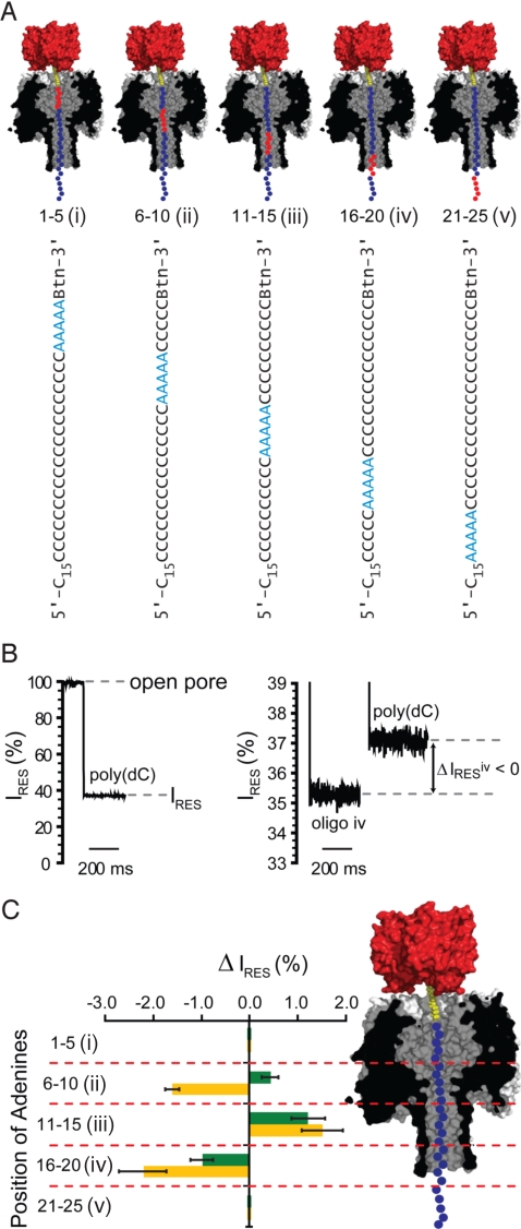
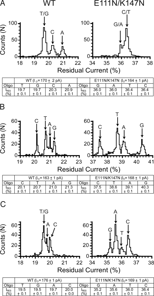
The sequencing of individual DNA strands with nanopores is under investigation as a rapid, low-cost platform in which bases are identified in order as the DNA strand is transported through a pore under an electrical potential. Although the preparation of solid-state nanopores is improving, biological nanopores, such as alpha-hemolysin (alphaHL), are advantageous because they can be precisely manipulated by genetic modification. Here, we show that the transmembrane beta-barrel of an engineered alphaHL pore contains 3 recognition sites that can be used to identify all 4 DNA bases in an immobilized single-stranded DNA molecule, whether they are located in an otherwise homopolymeric DNA strand or in a heteropolymeric strand. The additional steps required to enable nanopore DNA sequencing are outlined.


Conflict of interest statement

Conflict of interest statement: Hagan Bayley is the Founder, a Director, and a shareholder of Oxford Nanopore Technologies, a company engaged in the development of nanopore sequencing technology. This article was not supported by Oxford Nanopore Technologies.

Figures

Fig. 1.
Fig. 1.
Discrimination of immobilized DNA homopolymers by αHL pores. (A) Schematic representation of a homopolymeric DNA oligonucleotide (blue circles, only the first 25 nucleotides of the 60-nucleotide-long sequence are shown) immobilized inside an αHL pore (gray, cross-section) through the use of a biotin (yellow)–streptavidin (red) linkage. The αHL pore can be divided into 2 halves, each ≈5 nm in length; an upper vestibule located between the cis entrance and the central constriction, and a 14-stranded, transmembrane, antiparallel β-barrel, located between the central constriction and trans exit. The central constriction of 1.4 nm diameter is formed by the Glu-111, Lys-147 (shaded green), and Met-113 side chains contributed by all 7 subunits. (B and C Left) Current levels for the WT and E111N/K147N pores when blocked with immobilized poly(dC) and poly(dA) oligonucleotides. (B and C Right) Typical event histograms displaying the residual current levels, caused by poly(dC) and poly(dA) oligonucleotide blockages, for the WT and E111N/K147N pores. The mean residual current levels for each oligonucleotide were determined by performing Gaussian fits to the data.
Fig. 2.
Fig. 2.
Probing DNA recognition by the αHL pore with A5 oligonucleotides. (A) The 5 oligonucleotides (i–v) containing 5 consecutive adenine nucleotides (A5, red circles) at different positions (numbered from the 3′-biotin tag) in an otherwise poly(dC) strand (cytidine nucleotides are shown as blue circles). Only the first 25 of the 40-nucleotide-long sequences are shown. (B Left) The stepwise reduction from the open current value (pore not blocked with DNA) to a residual current (IRES) level of ≈37% when the E111N/K147N pore becomes blocked with a poly(dC) oligonucleotide. (B Right) The IRES levels when a pore is blocked with oligonucleotides of different sequence (oligo iv and poly(dC) are shown). (C) Residual current difference (ΔIRES) between the blockade by oligonucleotides i–v (A) and poly(dC)40 for WT (green bars) and E111N/K147N (orange bars) αHL pores (ΔIRES = IRESi–vIRESpoly(dC)). The probable location of the adenine (A5) stretch of each oligonucleotide when immobilized with an αHL pore is indicated (Right).
Fig. 3.
Fig. 3.
Discrimination of a single adenine nucleotide by αHL. The graph (Middle) indicates the differences in residual current (ΔIRES values) between blockades caused by a poly(dC) oligonucleotide containing a single adenine nucleotide (the sequence of each oligonucleotide is shown to the left) and blockades caused by poly(dC)40 for WT (green) and E111N/K147N (orange) αHL pores (see also Table S4). R1, R2 and R3 represent the 3 proposed recognition sites in the αHL nanopore. Their probable locations are indicated on the cross-section of the β barrel domain of the αHL pore (Right).
Fig. 4.
Fig. 4.
Recognition of all 4 DNA bases sites by the WT and E111N/K147N αHL pores. Histograms of the residual current levels for WT (Left) and E111N/K147N (Right) pores are shown. Three sets of 4 poly(dC) oligonucleotides were used, with each set containing either a single G, A, T, or C nucleotide at a specific position. All experiments were conducted at least 3 times, and the results displayed in the figure are from a typical experiment. (A) The WT and E111N/K147N pores were interrogated with 5′-CCCCCCCCCCCCCCCCCCCCCCCCCCCCCCCNCCCCCCCCBtn-3′ where N represents either G, A, T, or C. Gaussian fits were performed for each peak, and the mean value of the residual current for each oligonucleotide (and the standard deviation) is displayed in the table below the histograms. (B) WT and E111N/K147N pores were interrogated with 4 oligonucleotides with the sequence 5′-CCCCCCCCCCCCCCCCCCCCCCCCCCNCCCCCCCCCCCCCBtn-3′. (C) WT and E111N/K147N pores were interrogated with 4 oligonucleotides with the sequence 5′-CCCCCCCCCCCCCCCCCCCCCCNCCCCCCCCCCCCCCCCCBtn-3′.
Fig. 5.
Fig. 5.
Probing the E111N/K147N αHL pore for single-nucleotide discrimination in a heteropolymeric oligonucleotide. Histogram (Top) of residual current levels for E111N/K147N pores interrogated with 4 heteropolymeric DNA strands (Middle) that differ at only 1 position (blue). Gaussian fits were performed for each peak, and the mean value of the residual current for each oligonucleotide (and the standard deviation) is displayed (Bottom).

References

    1. Schloss JA. How to get genomes at one ten-thousandth the cost. Nat Biotechnol. 2008;26:1113–1115. - PubMed
    1. Kahvejian A, Quackenbush J, Thompson JF. What would you do if you could sequence everything? Nat Biotechnol. 2008;26:1125–1133. - PMC - PubMed
    1. Mardis ER. Next-generation DNA sequencing methods. Annu Rev Genomics Hum Genet. 2008;9:387–402. - PubMed
    1. Bayley H. Sequencing single molecules of DNA. Curr Opin Chem Biol. 2006;10:628–637. - PubMed
    1. Gupta PK. Single-molecule DNA sequencing technologies for future genomics research. Trends Biotechnol. 2008;26:602–611. - PubMed

Publication types

LinkOut - more resources