. 2009 Apr 20;185(2):279-90.
doi: 10.1083/jcb.200809153.

Bax activation by the BH3-only protein Puma promotes cell dependence on antiapoptotic Bcl-2 family members

Affiliations

Bax activation by the BH3-only protein Puma promotes cell dependence on antiapoptotic Bcl-2 family members

Tristan Gallenne et al. J Cell Biol. .

Abstract

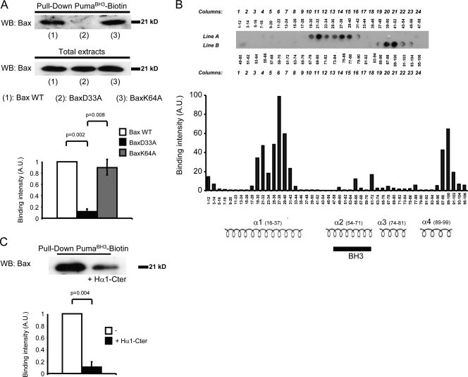
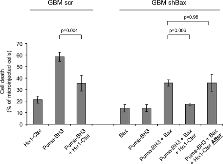
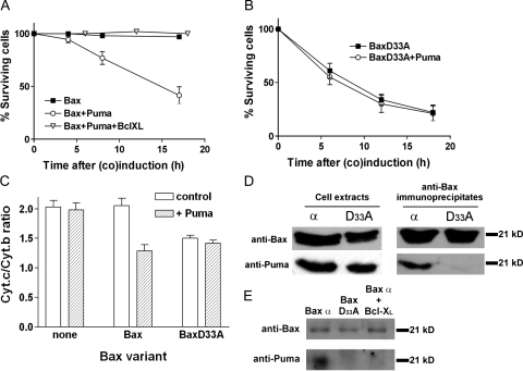
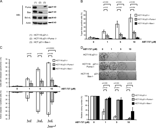
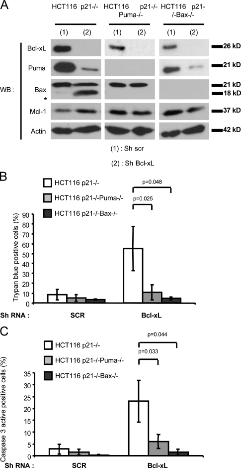
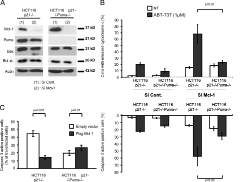
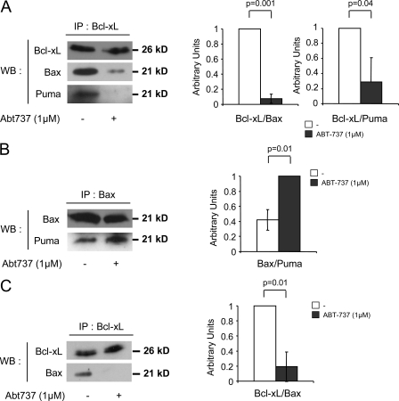
It is still unclear whether the BH3-only protein Puma (p53 up-regulated modulator of apoptosis) can prime cells to death and render antiapoptotic BH3-binding Bcl-2 homologues necessary for survival through its ability to directly interact with proapoptotic Bax and activate it. In this study, we provide further evidence, using cell-free assays, that the BH3 domain of Puma binds Bax at an activation site that comprises the first helix of Bax. We also show that, in yeast, Puma interacts with Bax and triggers its killing activity when Bcl-2 homologues are absent but not when Bcl-xL is expressed. Finally, endogenous Puma is involved in the apoptotic response of human colorectal cancer cells to the Bcl-2/Bcl-xL inhibitor ABT-737, even in conditions where the expression of Mcl-1 is down-regulated. Thus, Puma is competent to trigger Bax activity by itself, thereby promoting cellular dependence on prosurvival Bcl-2 family members.


Figures

Figure 1.
Figure 1.
Role of the C-terminal end of Bax Hα1 in the interaction of Bax with the BH3 domain of Puma. (A) Interaction of cellular Bax with a Puma BH3 peptide. Lysates from BdGBM(Bax-α), BdGBM(BaxD33A), and BdGBM(BaxK64A) cells expressing comparable amounts of Bax were used. Bax molecules pulled down from these lysates by biotinylated PumaBH3 were evaluated by Western blotting. (bottom) In each independent experiment, the amount of pulled down Bax was quantitated by densitometry and normalized to the maximum intensity. (B) Pepscan analysis. Interaction of biotinylated PumaBH3 with nitrocellulose membranes, on which were spotted 48 12-mer peptides covering the indicated sequence of Bax, was analyzed as described in Materials and methods. The top panel shows one representative autoradiogram. Peptides range from A1, which is the 12-mer peptide encompassing residues 1–12, to B24, which is the 12-mer peptide encompassing residues 95–106. In the bottom panel, the mean binding intensities (which were evaluated by densitometry and normalized to the maximum binding intensity) for three independent experiments using three different membranes are represented for each peptide. The positions of helices 1–4 (as defined in Suzuki et al. [2000]) are shown for illustration. (C) Interaction of cellular Bax with PumaBH3 in the presence of Bax Hα1-Cter peptide. Pull-down experiments were performed as in A, using lysates from BeGBM cells. Where indicated, 10 µM Bax Hα1-Cter was added to lysates before pull-down. (A and C) Data are mean ± SEM of three independent experiments. P-values were assessed using a Student's t test. A.U., arbitrary unit; WB, Western blot; WT, wild type.
Figure 2.
Figure 2.
Functional cooperation between PumaBH3 and Bax. The indicated GBM cells were microinjected with combinations of 100 nM PumaBH3, 1 µM Hα1-Cter, and/or 100 nM recombinant Bax together with a fluorescent microinjection marker (0.5% FITC–dextran 40S). Where indicated, Hα1-Cter was incubated with PumaBH3 before incubation with Bax or after PumaBH3 was incubated with Bax. The percentage of properly microinjected (i.e., fluorescent) cells exhibiting morphological features of cell death was assayed 6 h after microinjection. Data are mean ± SEM of at least three independent experiments. P-values were assessed using a Student's t test.
Figure 3.
Figure 3.
Interaction of Bax and Puma in yeast. (A) Yeast cell survival after Bax, Puma, and Bcl-xL coexpression. Induction of Bax, Puma, and/or Bcl-xL in the corresponding yeast cells and survival assays at the indicated time after induction were performed as described in Materials and methods. (B) Yeast cell survival after BaxD33A and Puma coexpression. The experiment was performed as in A. (C) Cytochrome c release after wild-type Bax, BaxD33A, and Puma coexpression. Cytochrome (Cyt.) content in mitochondria isolated from the indicated yeast cells 14 h after induction was measured as described in Materials and methods. (D) Puma interaction with wild-type Bax and BaxD33A in yeast. Extracts from cells coexpressing Bax (or BaxD33A) and Puma were immunoprecipitated with anti-Bax antibody, and immunoprecipitates were analyzed for the presence of Bax and Puma by Western blotting. (E) Effect of Bcl-xL on Puma–Bax interactions in yeast. Extracts from cells coexpressing Bax and Puma, BaxD33A and Puma, or Bax, Puma, and Bcl-xL were treated as in D. (A–C) Data are mean ± SEM of three independent experiments.
Figure 4.
Figure 4.
Role of endogenous Puma in the biological activity of ABT-737. (A) Expression of Bcl-2 family members in HCT116-derived cell lines. Expression of 50 µg Puma, Bax, and Bcl-xL in lysates from the indicated cells was analyzed by Western blotting. WB, Western blot. (B) Induction of cell death by ABT-737 in human colorectal cancer cells. The indicated cells were incubated with the indicated concentration of ABT-737 for 24 h, and induction of cell death was assayed by a trypan blue–staining procedure. Data are mean ± SEM of four independent experiments. (C) Induction of apoptosis by ABT-737 in human colorectal cancer cells. The indicated cells were incubated with the indicated concentration of ABT-737 for 24 h before immunocytochemical analysis of the percentage of cells exhibiting cytochrome c release and caspase 3 activation. Data are mean ± SEM of three independent experiments. (D) Long-term effects of ABT-737 on human colorectal cells. The indicated cells were treated for 2 wk with the indicated concentration of ABT-737 before analysis of viable clones by crystal violet staining. Data are mean ± SEM of at least three independent experiments and are expressed as a percentage of the number of colonies formed in untreated dishes. One experiment representative of three independent ones is shown in the top panel. (B–D) P-values were assessed using a Student's t test.
Figure 5.
Figure 5.
Role of endogenous Puma in the induction of apoptotic cell death by down-regulation of Bcl-xL in human cancer colorectal cells. (A) Expression of Bcl-2 family members in HCT116-derived cell lines after down-regulation of Bcl-xL. Western blot (WB) analysis of Puma, Bax, Bcl-xL, and Mcl-1 in lysates from the indicated cells was performed 3 d after infection with the indicated recombinant lentivirus. The asterisk indicates a truncated form of Bax of 18 kD. (B) Induction of cell death by down-regulation of Bcl-xL in human colorectal cancer cells. The indicated cells were infected with the indicated lentivirus, and induction of cell death was assayed by a trypan blue–staining procedure 3 d later. (C) Activation of caspase 3 by down-regulation of Bcl-xL in human colorectal cancer cells. The indicated cells were infected with the indicated recombinant lentivirus, and activation of caspase 3 was evaluated as described in Fig. 4 B 2 d later. (B and C) Data are mean ± SEM of three independent experiments. P-values were assessed using paired Student's t tests. ShRNA, short hairpin RNA.
Figure 6.
Figure 6.
Role of Mcl-1 in Puma-dependent apoptosis induced by ABT-737. (A and B) Down-regulation of Mcl-1. The indicated cells were transfected with the indicated siRNA. 48 h later, Western blot analysis of Mcl-1, Puma, Bax, and Bcl-xL (A) and treatment with 1 µM ABT-737 (B) were performed. Induction of cytochrome c release and caspase 3 activation were analyzed as described for Fig. 4 B. NT, not treated. (C) Overexpression of Mcl-1. The indicated cells were transfected with the indicated plasmid. 24 h later, cells were treated with 10 µM ABT-737 for an additional 24 h. Immunocytochemical analysis of active caspase 3 and of Flag expression was then performed. For Flag–Mcl-1–transfected cells, the percentage of Flag-positive cells that also stained positive for active caspase 3 was evaluated. For mock-transfected cells, the percentage of cells that stained positive for active caspase 3 was evaluated in random populations. (B and C) Data are mean ± SEM of three independent experiments. P-values were assessed using a Student's t test.
Figure 7.
Figure 7.
Effect of ABT-737 on intermolecular interactions involving Bax, Bcl-xL, and Puma. (A) Interactions between Bcl-xL and Bax or Puma. HCT116 p21−/− cells were treated or not treated (−) with 1 µM ABT-737 for 24 h. Cell lysates were immunoprecipitated (IP) with an anti–Bcl-xL antibody as described in Materials and methods, and the presence of Bcl-xL, Bax, and Puma in the immunoprecipitated fractions was analyzed by immunoblotting. The amount of Bax and Puma that were coimmunoprecipitated with Bcl-xL in each condition was evaluated by densitometric analysis and normalized to the amount of protein that coimmunoprecipitated with Bcl-xL in untreated cells. (B) Interactions between Puma and Bax. Experiments were performed as in A using an anti-Bax antibody for immunoprecipitation. The amount of Puma that was coimmunoprecipitated with Bax in each condition (evaluated by densitometric analysis) was normalized to the amount of protein that coimmunoprecipitated with Bax in treated cells. (C) Interactions between Bcl-xL and Bax in Puma knockout cells. Experiments were performed as in A using HCT116 p21−/−Puma−/− cells. (A–C) Data are mean ± SEM of three independent experiments. P-values were assessed using a Student's t test. WB, Western blot.

Comment in

  • Puma strikes Bax.
    Letai A. Letai A. J Cell Biol. 2009 Apr 20;185(2):189-91. doi: 10.1083/jcb.200903134. J Cell Biol. 2009. PMID: 19380876 Free PMC article.

References

    1. Adams J.M., Cory S. 2007. The Bcl-2 apoptotic switch in cancer development and therapy.Oncogene. 26:1324–1337 - PMC - PubMed
    1. Arokium H., Camougrand N., Vallette F.M., Manon S. 2004. Studies of the interaction of substituted mutants of BAX with yeast mitochondria reveal that the C-terminal hydrophobic alpha-helix is a second ART sequence and plays a role in the interaction with anti-apoptotic BCL-xL.J. Biol. Chem. 279:52566–52573 - PubMed
    1. Arokium H., Ouerfelli H., Velours G., Camougrand N., Vallette F.M., Manon S. 2007. Substitutions of potentially phosphorylatable serine residues of Bax reveal how they may regulate its interaction with mitochondria.J. Biol. Chem. 282:35104–35112 - PubMed
    1. Bellot G., Cartron P.F., Er E., Oliver L., Juin P., Armstrong L.C., Bornstein P., Mihara K., Manon S., Vallette F.M. 2007. TOM22, a core component of the mitochondria outer membrane protein translocation pore, is a mitochondrial receptor for the proapoptotic protein Bax.Cell Death Differ. 14:785–794 - PubMed
    1. Brix J., Rudiger S., Bukau B., Schneider-Mergener J., Pfanner N. 1999. Distribution of binding sequences for the mitochondrial import receptors Tom20, Tom22, and Tom70 in a presequence-carrying preprotein and a non-cleavable preprotein.J. Biol. Chem. 274:16522–16530 - PubMed

Publication types

MeSH terms