. 2009 Apr 20;185(2):323-39.
doi: 10.1083/jcb.200809046.

Defining mechanisms of actin polymerization and depolymerization during dendritic spine morphogenesis

Affiliations

Defining mechanisms of actin polymerization and depolymerization during dendritic spine morphogenesis

Pirta Hotulainen et al. J Cell Biol. .

Abstract

Dendritic spines are small protrusions along dendrites where the postsynaptic components of most excitatory synapses reside in the mature brain. Morphological changes in these actin-rich structures are associated with learning and memory formation. Despite the pivotal role of the actin cytoskeleton in spine morphogenesis, little is known about the mechanisms regulating actin filament polymerization and depolymerization in dendritic spines. We show that the filopodia-like precursors of dendritic spines elongate through actin polymerization at both the filopodia tip and root. The small GTPase Rif and its effector mDia2 formin play a central role in regulating actin dynamics during filopodia elongation. Actin filament nucleation through the Arp2/3 complex subsequently promotes spine head expansion, and ADF/cofilin-induced actin filament disassembly is required to maintain proper spine length and morphology. Finally, we show that perturbation of these key steps in actin dynamics results in altered synaptic transmission.


Figures

Figure 1.
Figure 1.
Sites of actin filament polymerization in dendritic filopodia. (A and B) The free actin filament barbed ends in mouse hippocampal neurons were visualized with fluorescently labeled actin monomers (middle panels). F-actin was stained with fluorescently labeled phalloidin (left panels). Right panels show merged images. The cell in A was fixed at DIV 16 and the cell in B was fixed at DIV 12. (C) The number of sites (percentage of total amount) of barbed ends in different locations (tip, root, or tip + root) was counted from 76 filopodia from four independent experiments (DIV 12–16). Data are presented as mean ± SEM. (D and E) Mouse cortical neurons were transfected with a construct expressing GFP–β-actin at DIV 8, and the FRAP assay was performed at DIV 9. GFP-actin was bleached from total filopodium (red), and the appearance of nonbleached actin (green) was followed by time-lapse imaging. In D, GFP-actin fluorescence recovers (actin filaments polymerize) from the filopodium tip; in E, recovery occurs mainly from the root. Bars, 1 µm.
Figure 2.
Figure 2.
Role of Rif in spinogenesis. (A) Mouse hippocampal neurons were transfected with GFP alone, or with GFP and myc-tagged inactive Rif or constitutively active Rif at DIV 11. Cells were fixed at DIV 12, and expression of Rif myc constructs was detected by anti-myc antibody (not depicted). Bars, 5 µm. (B) Quantitative analysis of neurons expressing inactive Rif (inact Rif) did not reveal significant changes in dendritic protrusion density as compared with wild-type cells (wt). Dendritic protrusion length was slightly reduced. Numerical data and p-values are presented in Table IV. (C) Dendritic protrusion morphology analysis of neurons expressing inactive Rif revealed a significant decrease in the number of thin spines and an increase in the number of stubby spines. Numerical data and p-values are presented in Tables I and II. (D) Quantitative dendritic protrusion analysis of neurons expressing active Rif (act Rif) revealed a significant reduction in dendritic protrusion density, whereas the mean length of dendritic protrusions was comparable to wild-type cells (wt). Numerical data are presented in Table IV. (E) Dendritic protrusion morphology analysis of neurons expressing active Rif did not reveal statistically significant changes between GFP and active Rif-transfected cells. Numerical data are shown in Tables I and II. Graphs represent mean ± SEM.
Figure 3.
Figure 3.
Role of mDia2 in spinogenesis. (A) Mouse hippocampal neurons were transfected with a plasmid expressing GFP (left) or a GFP fusion of active mDia2 construct at DIV 11, then fixed and stained with phalloidin at DIV 12. Active mDia2 localized to the filopodia tips, and its expression induced filopodia and spine head loss. (B) Mouse hippocampal neurons were transfected with GFP (left) or GFP-active mDia2 (green) and inactive Rif-myc (red) constructs at DIV 12; the cells were fixed and stained with anti-myc antibodies at DIV 13. Expression of active mDia2 overcomes the effect of inactive Rif. Arrows indicate the GFP-mDia2 tip localization. (C) Quantitative analysis of neurons expressing inactive Rif and active mDia2 (inactRif + actmDia2) showed significant reduction in dendritic protrusion density and length as compared with wild-type (wt) cells. Numerical data and p-values are presented in Table IV. (D) Dendritic protrusion morphology analysis of inactive Rif and active mDia2-expressing neurons revealed an increase in the number of filopodia and a significant decrease in the number of thin spines. Numerical data and p-values are presented in Tables I and II. (E) Mouse hippocampal neurons were transfected with GFP-actin, GFP-actin + mDia2 siRNA, or GFP-actin + inactive Rif constructs at DIV 12; the cells were fixed and stained with anti-mDia2 antibodies (Fig. S3) or anti-myc antibodies (not depicted) at DIV 13. Transfection of cells with mDia2 siRNA oligonucleotides resulted in dendritic protrusion morphology defects similar to those from expression of inactive Rif (shortened spine necks and larger spine heads). (F) Quantitative analysis of mDia2 siRNA-treated neurons showed a significant reduction in dendritic protrusion density and dendritic protrusion length as compared with wild type (wt). Numerical data and p-values are presented in Table IV. (G) Dendritic protrusion morphology analysis of mDia2 siRNA-treated neurons revealed a decrease in the number of filopodia and thin spines, and a significant increase in the number of stubby spines. Numerical data and p-values are presented in Tables I and II. Graphs represent mean ± SEM. Bars, 5 µm.
Figure 4.
Figure 4.
Identification of the sites of actin filament polymerization in spine heads. (A–C) The free actin filament barbed ends in mouse hippocampal neurons (DIV 12) were visualized with fluorescently labeled actin monomers (middle panels, barbed ends are indicated with arrowheads). F-actin was stained with fluorescently labeled phalloidin (left panels). Right panels show merged pictures. Barbed ends localized either as dots (A and C) or a “line” (B) at the spine head surface or to the root of the neck (C). (D) The number of sites (percentage of total spines analyzed) of barbed ends in different locations (tip of spine head [head tip] or tip of spine head + root of the neck [h. tip + n. root]) were counted from 128 spines from four independent experiments (DIV 12–16). The graph represents mean ± SEM. (E) Mouse hippocampal neurons were transfected with GFP-actin at DIV 20, and the FRAP assay was performed at DIV 21. GFP-actin was bleached from the spine head, and recovery of the GFP-actin fluorescence was followed by time-lapse imaging. The fluorescence of GFP-actin recovers mainly from the spine head tip. First sites of recovery are indicated with arrowheads. Bars, 1 µm.
Figure 5.
Figure 5.
Arp2/3 is necessary for spine head formation. (A) Mouse hippocampal neurons were transfected with GFP or GFP + p34 siRNA at DIV 10. At DIV 11, the cells were fixed and stained with anti-p34 antibodies (see Fig. S4). Reduced p34 levels resulted in a loss of spine heads. Bars, 5 µm. (B) Dendritic protrusion density and length of p34 siRNA-transfected neurons was analyzed with NeuronIQ software (Cheng et al., 2007). Dendritic protrusion density was decreased and dendritic protrusion length increased in p34 siRNA-transfected cells compared with wild-type cells. See Tables I and II for numerical data. (C) Dendritic protrusion morphology analysis revealed a clear reduction of thin, mushroom, and stubby spines. See Tables I and II for numerical data. (D) Mouse hippocampal neurons were transfected with GFP (wt) or inactive Rif + Scar1-WA (inact Rif + WA) constructs at DIV 10. At DIV 11, cells were fixed and stained with anti-myc antibodies. Inhibition of Arp2/3 and Rif induced spine head loss. Bars, 5 µm. (E) Dendritic protrusion density was reduced, whereas mean dendritic protrusion length was not affected in cells expressing inactive Rif and Scar1-WA. See Table IV for numerical data. (F) Morphology analysis revealed a significant reduction in thin, mushroom, and stubby spines in cells expressing inactive Rif and Scar1-WA. See Tables I and II for numerical data. Graphs represent mean ± SEM.
Figure 6.
Figure 6.
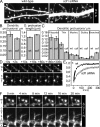
Cofilin-1 is necessary for proper spine morphology and actin turnover. (A) Mouse hippocampal neurons were transfected with GFP in the presence or absence of cofilin-1 siRNA oligonucleotides at DIV 11. On DIV 12, the cells were fixed and stained with anti–cofilin-1 antibodies (see Fig. S5). Reduction in cofilin-1 protein levels resulted in abnormal spine morphology (longer necks, branched spine heads). (B) Dendritic protrusions of wild-type and cofilin-1 siRNA-transfected cells were analyzed with NeuronIQ software (Cheng et al., 2007). Dendritic protrusion density was reduced and dendritic protrusion length was increased in cofilin-1 siRNA-transfected cells as compared with wild-type cells. Numerical data are presented in Table IV. (C) Morphology analysis revealed a decrease in the number of thin spines. Numerical data are presented in Tables I and II. Moreover, the density of branched spines was increased from 0.015 to 0.039 spines per micrometer (P < 0.001) as compared with wild-type cells. Graphs represent mean ± SEM. (D) The rate of actin turnover was analyzed from wild-type and cofilin-1 siRNA-transfected cells by FRAP. Mouse cortical neurons were transfected with GFP-actin (wt) or with GFP-actin with cofilin-1 siRNA (cof) at DIV 11, and the FRAP analysis was performed at DIV 12. The frames before (−10 s) and after bleach (from +2 to +306 s) are shown. In the spines of wild-type neurons, the fluorescence of GFP-actin recovery was nearly complete at 80 s; in cofilin-1 siRNA neurons, complete recovery was not achieved within 306 s. (E) The averaged recovery curves from nine wild-type and seven cofilin-1 siRNA cells revealed a diminished rate of actin turnover in cofilin-1 siRNA-transfected cells. Error bars represent SEMs. (F) Dynamics of spines of wild-type and cofilin-1 siRNA-transfected cells were followed by time-lapse stack confocal scan microscopy. Mouse hippocampal neurons at DIV 11 (wild-type) or DIV 10 (cofilin-1 siRNA) were transfected with GFP-actin without (wt; Video 7) or with cofilin-1 siRNA oligonucleotides (cofsiRNA; Video 9), and the time-lapse videos were acquired on the day after the transfection. Note the slow removal of the filopodia-like protrusions from the spine head in the cofilin-1–depleted cell. Bars, 1 µm.
Figure 7.
Figure 7.
Manipulation of the actin cytoskeleton affects the number of synapses and frequency of mEPSCs. (A) Mouse hippocampal neurons were transfected with GFP + control siRNA oligonucleotides (wt), with a Scar1-WA construct (inhibits Arp2/3; WA), with an inactive Rif construct (Rif), or with GFP + cofilin-1 siRNA (cof) at DIV 11. At DIV 12, the cells were fixed and stained with anti–V-GLUT-1 antibodies (green) to label the presynaptic part of synapses. In addition, the cells expressing myc-tagged constructs, WA and Rif, were stained with anti-myc antibodies (red, as in GFP staining). Bars, 5 µm. (B) The number of synapses was counted with Imaris software from at least 10 cells from each group. The WA expression reduced the number of synapses from 0.27 to 0.24 per micrometer; P = 0.023. Rif expression or cofilin-1 siRNA transfections caused only a slight reduction to 0.26 per micrometer (P = 0.326) or 0.25 per micrometer (P = 0.122), respectively. The graph indicates SEMs. (C) Functional synapses were analyzed by measuring mEPSCs. Mouse hippocampal neurons were transfected with GFP alone (wild-type) or with GFP with p34 siRNA, with an inactive Rif construct, or with cofilin-1 siRNA. siRNA was transfected at DIV 11, and plasmids were transfected at DIV 13. Shown are representative recordings of spontaneous miniature glutamatergic postsynaptic currents of the neurons at DIV 14. (D) The cumulative probability plots from the recordings presented in C show an increased appearance of longer inter-event intervals for transfected cells but no obvious changes in the event amplitudes. (E) Diagram showing the overall effect of transfection with p34 or cofilin-1 siRNA or an inactive Rif construct on inter-event interval and event amplitude. Median inter-event intervals or amplitude of each manipulated cell have been divided by the wild-type values of the same experimental day (DIV 11–14). The mean time between events was twice as long in p34 siRNA (n = 9 experimental days, 27 cells, P = 0.009) or inactive Rif-transfected cells (n = 8 experimental days, 24 cells, P = 0.006) compared with control cells (43 cells). In cofilin-1 siRNA cells (n = 9 experimental days, 27 cells, P = 0.421), the corresponding increase was 1.4. The amplitude of currents did not change with any manipulation. The graphs indicate SEMs.
Figure 8.
Figure 8.
A working model for the mechanisms of actin dynamics during dendritic spine development. (1) Spine development starts with the initiation of the dendritic filopodium and its elongation. At this stage, the filopodia are highly dynamic, undergoing continuous elongation and shrinking. We propose that mDia2 promotes actin filament polymerization in the filopodium tip. The factors driving actin filament polymerization in the roots of filopodia remains to be identified. (2) The spine head begins to form. We propose that the mechanism of actin assembly is gradually changed from an mDia2-mediated polymerization of unbranched actin filaments to an Arp2/3-nucleated branched actin filament network, leading to enlargement of the spine head. The spine heads are highly dynamic, continuously changing their shape, and long protrusions from the spine heads are frequently seen. We propose that the function of ADF/cofilins, in addition to replenishing the cytoplasmic actin monomer pool in neurons, is to control the proper length of actin filaments and thus to prevent formation of abnormal protrusions from spine heads. Future studies will be required to reveal the exact spine locations where ADF/cofilins are active. (3) Mature spines are still dynamic but maintain their overall morphology. Dynamics occur as small protrusions on the surface of the spine head (morphing).

Similar articles

Cited by

References

    1. Aoto J., Ting P., Maghsoodi B., Xu N., Henkemeyer M., Chen L. 2007. Postsynaptic EphrinB3 promotes shaft glutamatergic synapse formation.J. Neurosci. 27:7508–7519 - PMC - PubMed
    1. Bamburg J.R. 1999. Proteins of the ADF/cofilin family: essential regulators of actin dynamics.Annu. Rev. Cell Dev. Biol. 15:185–230 - PubMed
    1. Beli P., Mascheroni D., Xu D., Innocenti M. 2008. WAVE and Arp2/3 jointly inhibit filopodium formation by entering into a complex with mDia2.Nat. Cell Biol. 10:849–857 - PubMed
    1. Bertling E., Hotulainen P., Mattila P.K., Matilainen T., Salminen M., Lappalainen P. 2004. Cyclase-associated protein 1 (CAP1) promotes cofilin-induced actin dynamics in mammalian nonmuscle cells.Mol. Biol. Cell. 15:2324–2334 - PMC - PubMed
    1. Block J., Stradal T.E., Hänisch J., Geffers R., Köstler S.A., Urban E., Small J.V., Rottner K., Faix J. 2008. Filopodia formation induced by active mDia2/Drf3.J. Microsc. 231:506–517 - PubMed

Publication types

MeSH terms