. 2009 May;119(5):1230-40.
doi: 10.1172/JCI38022. Epub 2009 Apr 20.

Requirement for Ca2+/calmodulin-dependent kinase II in the transition from pressure overload-induced cardiac hypertrophy to heart failure in mice

Affiliations

Requirement for Ca2+/calmodulin-dependent kinase II in the transition from pressure overload-induced cardiac hypertrophy to heart failure in mice

Haiyun Ling et al. J Clin Invest. 2009 May.

Erratum in

  • J Clin Invest. 2012 Apr 2;122(4):1584. Heller Brown, Joan [corrected to Brown, Joan Heller]

Abstract

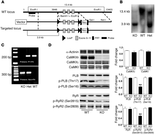
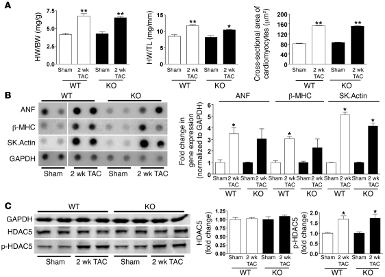
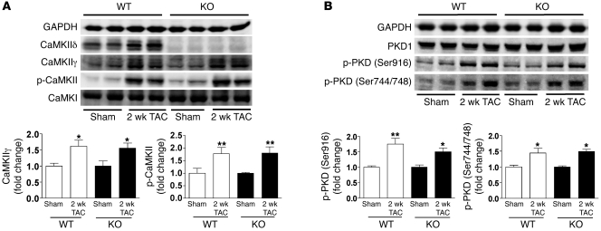
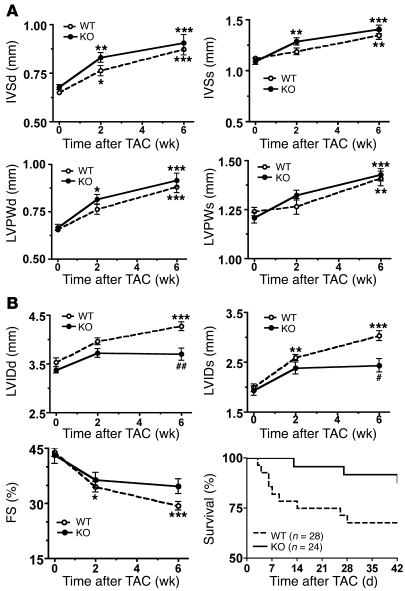
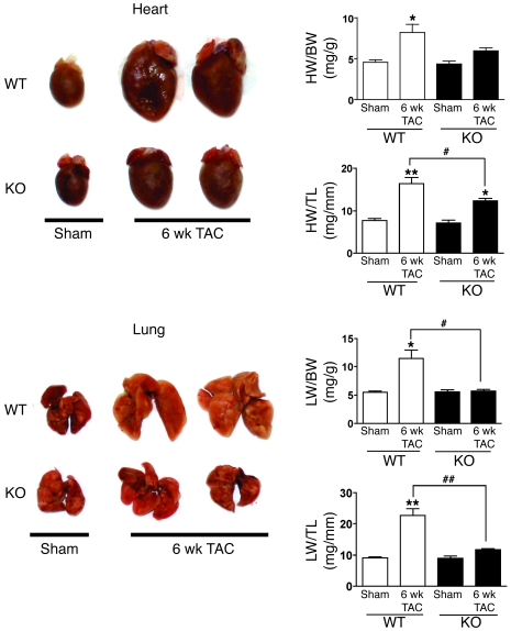
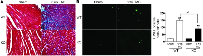
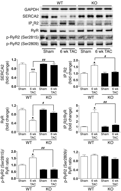
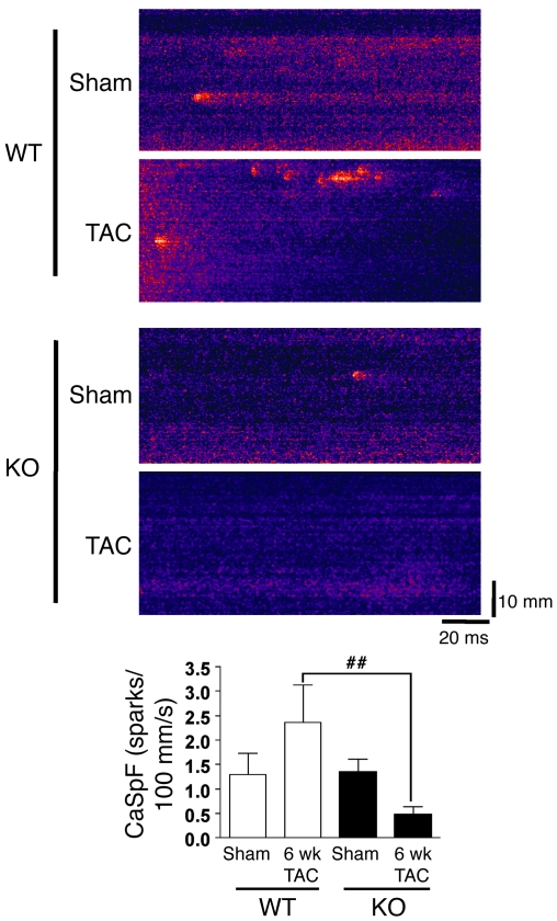
Ca2+/calmodulin-dependent kinase II (CaMKII) has been implicated in cardiac hypertrophy and heart failure. We generated mice in which the predominant cardiac isoform, CaMKIIdelta, was genetically deleted (KO mice), and found that these mice showed no gross baseline changes in ventricular structure or function. In WT and KO mice, transverse aortic constriction (TAC) induced comparable increases in relative heart weight, cell size, HDAC5 phosphorylation, and hypertrophic gene expression. Strikingly, while KO mice showed preserved hypertrophy after 6-week TAC, CaMKIIdelta deficiency significantly ameliorated phenotypic changes associated with the transition to heart failure, such as chamber dilation, ventricular dysfunction, lung edema, cardiac fibrosis, and apoptosis. The ratio of IP3R2 to ryanodine receptor 2 (RyR2) and the fraction of RyR2 phosphorylated at the CaMKII site increased significantly during development of heart failure in WT mice, but not KO mice, and this was associated with enhanced Ca2+ spark frequency only in WT mice. We suggest that CaMKIIdelta contributes to cardiac decompensation by enhancing RyR2-mediated sarcoplasmic reticulum Ca2+ leak and that attenuating CaMKIIdelta activation can limit the progression to heart failure.


Figures

Figure 1
Figure 1. Generation and characterization of mice with CaMKIIδ deletion.
(A) Restriction map of the genomic structure of the Camk2d gene, the targeting construct, and the mutated locus after recombination. The targeting construct was generated by flanking exons 9–11 of Camk2d with 2 loxP sites and flanking the Neo cassette by Flp recombinase target (FRT) sites. (B) Genotyping of CaMKIIδ-deficient mice by Southern blot analysis with the probe shown in A. Genomic DNA was isolated from the tails of a WT, a KO, and a heterozygous (Het) mouse. The 13.4- and 3.9-kb bands represent WT and mutant alleles, respectively. (C) Genotyping of KO mice by PCR analysis using mouse tail DNA and specific primers for the WT (P1+P2) and mutant (Neo+P4) alleles. (D) Analysis of protein expression and phosphorylation in WT and CaMKIIδ-deficient mice. LV homogenates were subjected to Western blotting. Quantitative analysis of the expression and phosphorylation of the proteins is shown at right. Data are mean ± SEM of 4–7 determinations. **P < 0.01 versus WT.
Figure 2
Figure 2. CaMKIIδ deletion does not prevent cardiac hypertrophy induced by 2-week TAC.
(A) Relative HW, indicated by HW/BW or HW/TL ratios, and cross-sectional area of cardiomyocytes detected by wheat germ agglutinin staining. Data are mean ± SEM of values from 3–6 mice. (B) Hypertrophic gene expression in response to 2-week TAC. RNA isolated from ventricular tissue of WT and KO mice was subjected to dot blot analysis using gene transcript–specific antisense oligonucleotide probes. GAPDH was used as the normalizing control. Samples for WT sham were loaded on the same membrane as the other but were noncontiguous (denoted by white line). Sk.Actin, α-skeletal actin. Data are mean ± SEM of values from 3 determinations. (C) Expression and phosphorylation of HDAC5 (Ser498) in LV homogenates after 2-week TAC, as detected by Western blot analysis. Data are mean ± SEM of values from 3–4 determinations. *P < 0.05, **P < 0.01 versus sham.
Figure 3
Figure 3. Upregulation of CaMKIIγ expression and PKD phosphorylation in cardiac hypertrophy.
(A) Expression of CaMKIIδ, CaMKIIγ, CaMKI, and phospho-CaMKII in LV homogenates 2 weeks after TAC, as detected by Western blot analysis. (B) Expression of PKD1 and phospho-PKD (Ser916 and Ser744/748) in LV homogenates 2 weeks after TAC were detected by Western blot analysis. Data are mean ± SEM of values from 4–6 determinations. *P < 0.05, **P < 0.01 versus sham.
Figure 4
Figure 4. Effects of CaMKIIδ deletion on the development of cardiac hypertrophy and heart failure after 6-week TAC.
Shown are averaged echocardiographic parameters and percent survival in WT and CaMKIIδ-deficient mice at different time points after TAC. Data are mean ± SEM of values from 12–18 mice, unless otherwise indicated. (A) Hypertrophic responses. (B) Decompensation to heart failure. Percent survival in response to TAC is plotted as a Kaplan-Meier curve (P = 0.08, WT versus KO). *P < 0.05, **P < 0.01, ***P < 0.001 versus pre-echocardiography; #P < 0.05, ##P < 0.01 versus WT.
Figure 5
Figure 5. CaMKIIδ deletion reduces relative HW and lung edema in response to 6-week TAC.
HW and LW were normalized to BW and TL. Data are mean ± SEM of values from 3–9 mice. *P < 0.05, **P < 0.01 versus sham; #P < 0.05, ##P < 0.01 versus WT TAC.
Figure 6
Figure 6. CaMKIIδ deletion inhibits cardiac fibrosis and apoptosis after 6-week TAC.
(A) Interstitial collagen deposition, indicative of cardiac fibrosis, as assessed by Masson trichrome staining. (B) TUNEL staining of heart tissue after 6-week TAC in WT and KO mice. Original magnification, ×40. Data are mean ± SEM of values from 3 hearts per group, with at least 5,000 nuclei examined per heart. **P < 0.01 versus sham; #P < 0.05 versus WT TAC.
Figure 7
Figure 7. CaMKIIδ deletion normalizes the expression of Ca2+ regulatory proteins and reduces RyR2 phosphorylation at the CaMKII site after 6-week TAC.
LV homogenates were subjected to Western blotting. Quantitative analysis is shown below. Phosphorylation of RyR2 at the CaMKII site (Ser2815) or at the PKA site (Ser2809) was normalized to RyR expression, and the IP3R2/RyR ratio was also calculated. Data are mean ± SEM of 3–4 determinations. *P < 0.05, **P < 0.01 versus sham; #P < 0.05, ##P < 0.01 versus WT TAC.
Figure 8
Figure 8. CaMKIIδ ablation reduces SR Ca2+ leak in 6-week TAC mice.
Longitudinal line scan images of spontaneous Ca2+ sparks were recorded in myocytes from both genotypes with or without TAC. CaSpF data were averaged from isolated cardiomyocytes in WT sham (n = 10), WT 6-week TAC (n = 7), KO sham (n = 12), and KO 6-week TAC (n = 12) groups. ##P < 0.01 versus WT TAC. There was no significant difference between KO and WT 6-week TAC mice with respect to SR Ca2+ content, assessed by caffeine-induced Ca2+ transient amplitude (fluorescence normalized to baseline, WT, 4.2 ± 0.5; KO, 3.8 ± 0.8).

Comment in

Similar articles

Cited by

References

    1. Bassani R.A., Mattiazzi A., Bers D.M. CaMKII is responsible for activity-dependent acceleration of relaxation in rat ventricular myocytes. Am. J. Physiol. 1995;268:H703–H712. - PubMed
    1. Hain J., Onoue H., Mayrleitner M., Fleischer S., Schindler H. Phosphorylation modulates the function of the calcium release channel of sarcoplasmic reticulum from cardiac muscle. J. Biol. Chem. 1995;270:2074–2081. doi: 10.1074/jbc.270.5.2074. - DOI - PubMed
    1. Karczewski P., Kuschel M., Baltas L.G., Bartel S., Krause E.G. Site-specific phosphorylation of a phospholamban peptide by cyclic nucleotide- and Ca2+/calmodulin-dependent protein kinases of cardiac sarcoplasmic reticulum. . Basic Res. Cardiol. 1997;92(Suppl. 1):37–43. - PubMed
    1. Witcher D.R., Kovacs R.J., Schulman H., Cefali D.C., Jones L.R. Unique phosphorylation site on the cardiac ryanodine receptor regulates calcium channel activity. J. Biol. Chem. 1991;266:11144–11152. - PubMed
    1. Wehrens X.H., Lehnart S.E., Reiken S.R., Marks A.R. Ca2+Calmodulin-dependent protein kinase ii phosphorylation regulates the cardiac ryanodine receptor. . Circ. Res. 2004;94:E61–E70. doi: 10.1161/01.RES.0000125626.33738.E2. - DOI - PubMed

Publication types

MeSH terms

Substances