. 2009 May;119(5):1167-77.
doi: 10.1172/JCI38482. Epub 2009 Apr 20.

Anti-TNF immunotherapy reduces CD8+ T cell-mediated antimicrobial activity against Mycobacterium tuberculosis in humans

Affiliations

Anti-TNF immunotherapy reduces CD8+ T cell-mediated antimicrobial activity against Mycobacterium tuberculosis in humans

Heiko Bruns et al. J Clin Invest. 2009 May.

Abstract

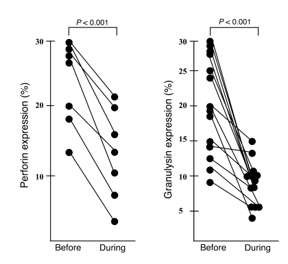
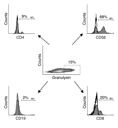
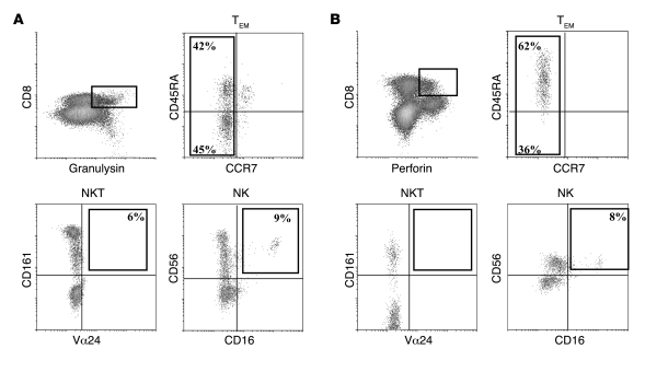
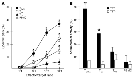
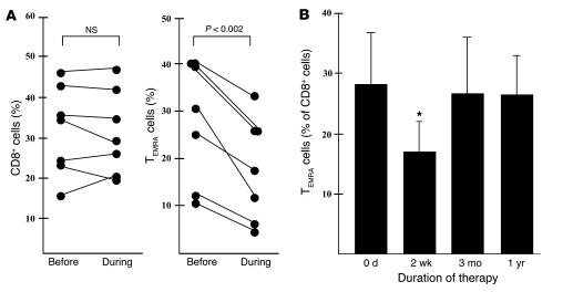
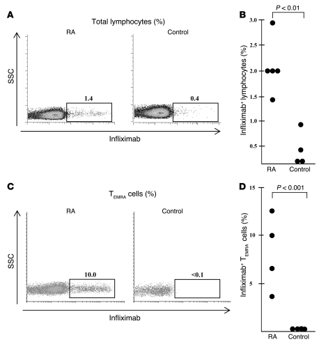
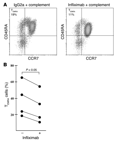
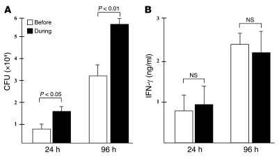
The incidence of tuberculosis is increased during treatment of autoimmune diseases with anti-TNF antibodies. This is a significant clinical complication, but also provides a unique model to study immune mechanisms in human tuberculosis. Given the key role for cell-mediated immunity in host defense against Mycobacterium tuberculosis, we hypothesized that anti-TNF treatment impairs T cell-directed antimicrobial activity. Anti-TNF therapy reduced the expression in lymphocytes of perforin and granulysin, 2 components of the T cell-mediated antimicrobial response to intracellular pathogens. Specifically, M. tuberculosis-reactive CD8+CCR7-CD45RA+ effector memory T cells (TEMRA cells) expressed the highest levels of granulysin, lysed M. tuberculosis, and infected macrophages and mediated an antimicrobial activity against intracellular M. tuberculosis. Furthermore, TEMRA cells expressed cell surface TNF and bound the anti-TNF therapeutic infliximab in vitro, making them susceptible to complement-mediated lysis. Immune therapy with anti-TNF was associated with reduced numbers of CD8+ TEMRA cells and decreased antimicrobial activity against M. tuberculosis, which could be rescued by the addition of CD8+ TEMRA cells. These results suggest that anti-TNF therapy triggers a reduction of CD8+ TEMRA cells with antimicrobial activity against M. tuberculosis, providing insight into the mechanism whereby key effector T cell subsets contribute to host defense against tuberculosis.


Figures

Figure 1
Figure 1. Decreased expression of lytic and antimicrobial effector molecules in patients treated with infliximab.
PBMCs from patients with active RA or AS were stained for perforin (n = 7) or granulysin (n = 17) before and 2 weeks after beginning therapy. The percentage of granulysin+ and perforin+ lymphocytes was determined by flow cytometry. Shown are results from all individual donors tested.
Figure 2
Figure 2. The majority of granulysin+ cells in the peripheral blood are cytotoxic lymphocytes.
PBMCs from healthy donors were stained with PE-Cy5-5–conjugated anti-CD4, PE-conjugated anti-CD56, allophycocyanin-conjugated anti-CD19, PerCP-conjugated anti-CD8 (filled histograms), or appropriate isotype controls (open histograms). All samples were then stained for granulysin using FITC-conjugated donkey anti-rabbit as a secondary antibody. Granulysin+ cells were gated according to the isotype (control rabbit serum, not shown) and analyzed for the expression of cell surface markers. The percentages of CD4+, CD56+, CD19+, and CD8+ cells within the granulysin gate are indicated. Shown is a typical result of 27 donors.
Figure 3
Figure 3. The majority of CD8+ and granulysin+ or perforin+ T cells are effector cells.
PBMCs from healthy donors were stained with PerCP- or allophycocyanin-conjugated anti-CD8 and anti-granulysin (detected with donkey anti-rabbit biotin and streptavidin) or PE-conjugated perforin. Additional labeling was performed using allophycocyanin-conjugated anti-CD45RA and FITC-conjugated anti-CCR7 to detect TEM cells, PE-conjugated anti-CD161 and FITC-conjugated anti-Vα24 to detect NKT cells, and PE-conjugated CD56 and FITC-conjugated anti-CD16 to detect NK cells. Samples with appropriate isotypes were included in all experiments. (A) CD8+granulysin+ cells were gated, and the expression of additional markers within this gate was determined. Numbers denote the percentage of positive effector cells, NKT cells, or NK cells within the population of CD8+granulysin+ cells. For each sample, 1 × 10⁶ cells were acquired. Shown is a typical result of 8 donors. (B) CD8+perforin+ cells were gated, and the expression of additional markers within this gate was determined. Numbers denote the percentage of positive effector cells, NKT cells, or NK cells within the population of CD8+perforin+ cells. For each sample, 1 × 10⁶ cells were acquired. Shown is a typical result of 4 donors.
Figure 4
Figure 4. CD8+ TEMRA cells lyse M. tuberculosis–infected monocytes and reduce mycobacterial growth.
CD8+ PBMCs were labeled with allophycocyanin-conjugated anti-CD45RA and FITC-conjugated anti-CCR7 and sorted into TEMRA, TEM, and TCM cell subpopulations. (A) Purified T cells were added as effector cells in a 51Cr release assay using autologous M. tuberculosis–infected (MOI 5) or uninfected monocytes as target cells. Supernatants were harvested after 4 hours, and 51Cr release was determined. Shown is mean ± SEM specific lysis of M. tuberculosis–infected monocytes of 4 independent experiments using different TST+ donors. *P < 0.05, infected versus uninfected monocytes. Lysis of uninfected monocytes was generally below 7%. (B) T cell subsets were purified as described above and incubated with infected autologous monocytes (5 × 10⁴ monocytes, 5 × 10⁵ T cells/well). After 36 hours, cells were lysed and plated in 10-fold dilutions on 7H9 agar plates. All samples were set up in duplicate. Shown is mean ± SEM reduction in the number of viable M. tuberculosis cells in monocytes cultured in the presence of T cells compared with monocytes cultured in the absence of T cells. The experiment was performed with 4 each of TST and TST+ donors. **P < 0.01 versus TST.
Figure 5
Figure 5. Decreased frequency of TEMRA cells in patients treated with infliximab.
PBMCs from patients with RA or AS were stained for CD8 (PE conjugated), CD45 (allophycocyanin conjugated), and CCR7 (FITC conjugated) before, 2 weeks, 3 months (n = 7), and 1 year (n = 5) after the beginning of infliximab therapy. (A) The percentage of all CD8+ T cells within the lymphocyte gate and the percentage of TEMRA cells within the CD8 gate were determined by flow cytometry. Shown is the percentage of CD8+ cells and TEMRA cells (n = 7) of all donors tested. (B) Mean number of TEMRA cells within CD8+ T cells of the same patients before and during infliximab therapy. *P < 0.05 versus pretreatment value.
Figure 6
Figure 6. PBMCs of RA patients express membrane TNF.
(A and B) PBMCs from patients with active RA (n = 5) or healthy controls (n = 4) were stained with biotinylated infliximab and allophycocyanin-conjugated streptavidin. Cells were acquired (1 × 10⁶) and analyzed by flow cytometry. (A) Percentage of infliximab+ lymphocytes of 1 representative patient (left panel) and healthy donor (right panel). (B) Individual results from all donors tested. (C and D) PBMCs from patients with active RA (n = 4) or healthy controls (n = 4) were stained with biotinylated infliximab, PerCP-conjugated streptavidin, PE-conjugated anti-CD8, allophycocyanin-conjugated anti-CD45RA, and FITC-conjugated anti-CCR7. CD8+ TEMRA cells were gated, and the percentage of infliximab+ cells was determined. For each sample, 5 × 10⁶ cells were acquired. (C) Percentage of CD8+infliximab+ TEMRA cells of 1 representative RA patient and healthy donor. (D) Individual results from all donors tested.
Figure 7
Figure 7. Complement reduces the frequency of infliximab+ TEMRA cells in RA patients.
PBMCs from patients with active RA (n = 4) were incubated with infliximab or a control antibody. Rabbit complement was then added for 60 minutes, and the number of TEMRA cells was determined by labeling with CD8, CD45RA, and CCR7. (A) CD8+ lymphocytes were gated, and the representative graph shows the CD45RA and CCR7 labeling within the CD8 gate. (B) Individual results of all donors investigated.
Figure 8
Figure 8. Infliximab therapy reduces the antimicrobial activity of PBMCs.
PBMCs from TST+ patients (n = 4) with active RA or AS were collected before and 2 weeks after the onset of anti-TNF therapy and frozen in liquid nitrogen to allow for simultaneous measurement of antimycobacterial activity. Matched pairs were thawed, and adherent monocytes were infected with M. tuberculosis at MOI 5. Infected monocytes were detached and plated in 24-well plates as described in Methods. Autologous nonadherent cells (1 × 10⁶) were added. (A) The number of viable bacilli was determined by plating the cell lysates at 24 and 96 hours after infection. Shown is the mean ± SEM number of CFUs of all 4 donors tested. (B) Supernatants from the same samples as in A were taken after 24 and 96 hours, and the concentration of IFN-γ was measured by ELISA. Shown is the mean ± SD concentration of IFN-γ of all 4 donors. The IFN-γ concentration in samples containing PBMCs, T cells, and uninfected macrophages was below the 30-pg/ml detection limit.
Figure 9
Figure 9. CD8+ TEMRA cells rescue antimycobacterial activity of PBMCs from patients during anti-TNF therapy.
PBMCs from 3 consecutive patients with active RA or AS and TST+ were collected before and 2 weeks after beginning of anti-TNF therapy. Adherent cells were infected with virulent M. tuberculosis at MOI 5 for 3 hours, and extracellular bacteria were removed. Of the macrophages, 21% ± 12% were infected (mean ± SD of 3 donors), with each macrophage harboring 1–3 bacilli. Infected monocytes were detached and plated in 24-well plates (5 × 10⁴). Concurrently, CD8+ T cells were purified from the nonadherent fraction and sorted by flow cytometry according to the CD45RA/CCR7 expression profile. CD20+ B cells were purified as a control population. Lymphocyte subsets were added to the infected monocytes (2 × 10⁵) as indicated, and the number of viable bacilli was determined by plating cell lysates after 36 hours of infection. Lysates were plated in 5-fold dilutions and in duplicates. The initial bacterial inoculum was 3.3 ± 0.7 × 10⁴. Shown is the mean ± SEM of 1 experiment with each individual donor. *P < 0.05 versus CFUs cultured in the presence of PBMCs during therapy.

Comment in

References

    1. Wolfe F., Michaud K., Anderson J., Urbansky K. Tuberculosis infection in patients with rheumatoid arthritis and the effect of infliximab therapy. Arthritis Rheum. 2004;50:372–379. doi: 10.1002/art.20009. - DOI - PubMed
    1. Elliott M.J., et al. Randomised double-blind comparison of chimeric monoclonal antibody to tumour necrosis factor alpha (cA2) versus placebo in rheumatoid arthritis. Lancet. 1994;344:1105–1110. doi: 10.1016/S0140-6736(94)90628-9. - DOI - PubMed
    1. Maini R., et al. Infliximab (chimeric anti-tumour necrosis factor alpha monoclonal antibody) versus placebo in rheumatoid arthritis patients receiving concomitant methotrexate: a randomised phase III trial. ATTRACT Study Group. Lancet. 1999;354:1932–1939. doi: 10.1016/S0140-6736(99)05246-0. - DOI - PubMed
    1. Feldmann M., Maini S.R. Role of cytokines in rheumatoid arthritis: an education in pathophysiology and therapeutics. Immunol. Rev. . 2008;223:7–19. doi: 10.1111/j.1600-065X.2008.00626.x. - DOI - PubMed
    1. Scott D.L., Kingsley G.H. Tumor necrosis factor inhibitors for rheumatoid arthritis. . N. Engl. J. Med. 2006;355:704–712. doi: 10.1056/NEJMct055183. - DOI - PubMed

Publication types

MeSH terms