. 2009 Aug 31;41(8):548-54.
doi: 10.3858/emm.2009.41.8.060.

Cordycepin inhibits UVB-induced matrix metalloproteinase expression by suppressing the NF-kappaB pathway in human dermal fibroblasts

Affiliations

Cordycepin inhibits UVB-induced matrix metalloproteinase expression by suppressing the NF-kappaB pathway in human dermal fibroblasts

Young Rae Lee et al. Exp Mol Med. .

Abstract

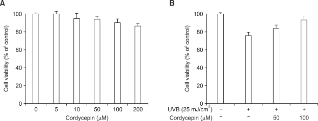
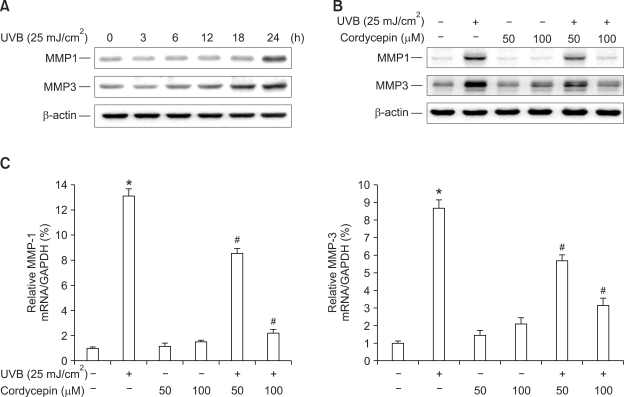
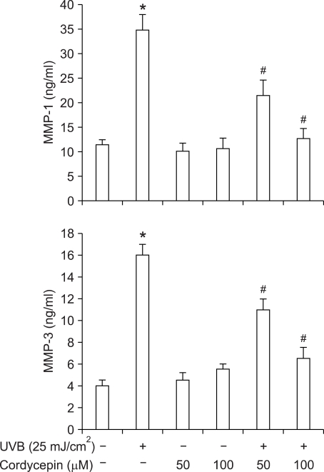
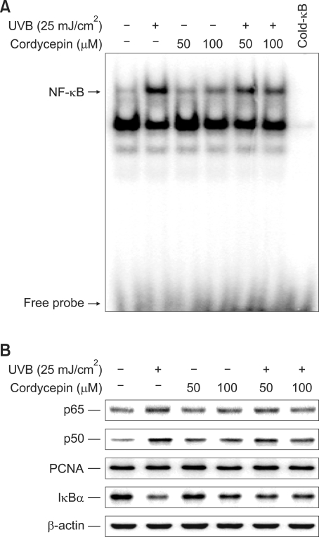
Cordycepin (3-deoxyadenosine) has been shown to exhibit many pharmacological activities, including anti-cancer, anti-inflammatory, and anti-infection activities. However, the anti-skin photoaging effects of cordycepin have not yet been reported. In the present study, we investigated the inhibitory effects of cordycepin on matrix metalloproteinase-1 (MMP-1) and -3 expressions of the human dermal fibroblast cells. Western blot analysis and real-time PCR revealed cordycepin inhibited UVB-induced MMP-1 and -3 expressions in a dose-dependent manner. UVB strongly activated NF-kappaB activity, which was determined by IkappaBalpha degradation, nuclear localization of p50 and p65 subunit, and NF-kappaB binding activity. However, UVB-induced NF-kappaB activation and MMP expression were completely blocked by cordycepin pretreatment. These findings suggest that cordycepin could prevent UVB-induced MMPs expressions through inhibition of NF-kappaB activation. In conclusion, cordycepin might be used as a potential agent for the prevention and treatment of skin photoaging.


Figures

Figure 1
Figure 1
Chemical structure of cordycepin.
Figure 2
Figure 2
Effect of cordycepin on cell viability in HDF. Cells were cultured in 96-well plates until 70% confluence, then incubated with the indicated concentration of cordycepin for 24 h. MTT assay was used to detect the viability of the cells as detailed in Methods (A). Cells were cultured in 100 mm culture dishes until 70% confluence, then incubated with the indicated concentration of cordycepin for 24 h in the presence of UVB. Cell viability determined by cell count method (B). The optical density value of control was regarded as 100%. Data points are the mean ± SE of three independent experiments.
Figure 3
Figure 3
Effect of cordycepin on UVB-induced MMP-1 and MMP-3 expressions in HDF. HDF in monolayer were incubated with UVB (25 mJ/cm2) in time dependent manner (A). To investigate effect of cordycepin, cells were stimulated with UVB (25 mJ/cm2) and the indicated concentrations of cordycepin for 24 h (B). The cell lysates were analyzed by Western blotting with anti-MMP-1 and -3. The blot was reprobed with anti-β-actin to confirm equal loading. HDF were pretreated with cordycepin for 1 h and stimulated by UVB (25 mJ/cm2) for 24 h. Total cellular RNA was analyzed by real-time PCR for MMP-1 and -3 (C). Each value represents the mean ± SEM of three independent experiments. *P < 0.01 vs. untreated control, #P < 0.01 vs. UVB.
Figure 4
Figure 4
Inhibition of UVB-induced MMP-1 and -3 secretions from HDF by cordycepin. Cells were stimulated with UVB (25 mJ/cm2) and the indicated concentrations of cordycepin for 24 h. Level of MMP-1 and -3 in culture media was measured using a commercially available ELISA kit as described in Methods. Each value represents the mean ± SEM of three independent experiments. *P < 0.01 vs. untreated control, #P < 0.01 vs. UVB.
Figure 5
Figure 5
Suppression of UVB-induced DNA binding of NF-κB, translocation of p65 and p50 to the nucleus, and IκBα degradation by cordycepin. Cells were stimulated with UVB (25 mJ/cm2) and the indicated concentrations of cordycepin. Following 3 h of incubation, DNA binding of NF-κB was analyzed by electrophoretic mobility shift analysis (A), and the translocation of p65 and p50 to the nucleus and IκBα degradation in the cytoplasm (B) were determined by Western blotting.

References

    1. Adhami VM, Afaq F, Ahmad N. Suppression of ultraviolet B exposure-mediated activation of NF-kappaB in normal human keratinocytes by resveratrol. Neoplasia. 2003;5:74–82. - PMC - PubMed
    1. Afaq F, Ahmad N, Mukhtar H. Suppression of UVB-induced phosphorylation of mitogen-activated protein kinases and nuclear factor kappa B by green tea polyphenol in SKH-1 hairless mice. Oncogene. 2003;22:9254–9264. - PubMed
    1. Bell S, Degitz K, Quirling M, Jilg N, Page S, Brand K. Involvement of NF-kappaB signalling in skin physiology and disease. Cell Signal. 2003;15:1–7. - PubMed
    1. Bond M, Baker AH, Newby AC. Nuclear factor kappaB activity is essential for matrix metalloproteinase-1 and -3 upregulation in rabbit dermal fibroblasts. Biochem Biophys Res Commun. 1999;264:561–567. - PubMed
    1. Bradford MM. A rapid, sensitive method for the quantitation of microgram quantities of protein utilizing the principle of protein-dye binding. Anal Biochem. 1976;72:248–254. - PubMed

Publication types

MeSH terms

LinkOut - more resources