. 2009;4(4):e5210.
doi: 10.1371/journal.pone.0005210. Epub 2009 Apr 21.

Phagocytosis of Staphylococcus aureus by macrophages exerts cytoprotective effects manifested by the upregulation of antiapoptotic factors

Affiliations

Phagocytosis of Staphylococcus aureus by macrophages exerts cytoprotective effects manifested by the upregulation of antiapoptotic factors

Joanna Koziel et al. PLoS One. 2009.

Abstract

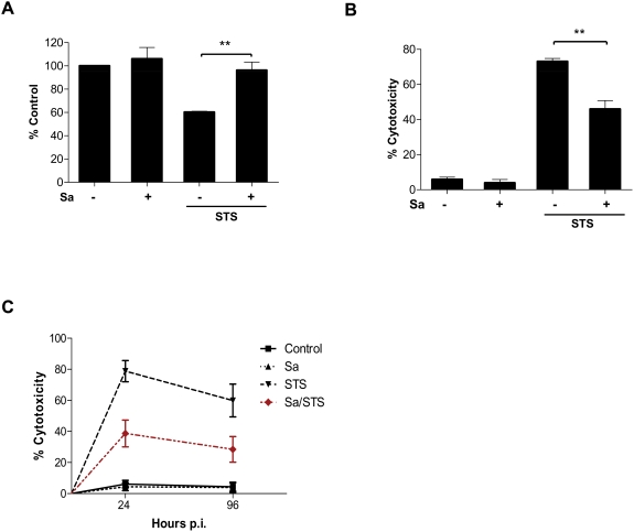
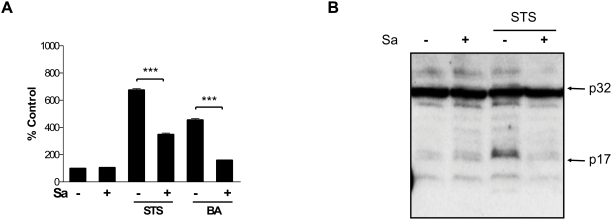
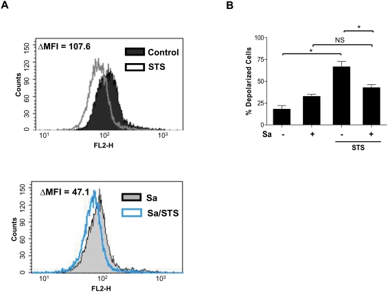
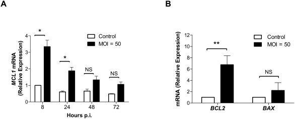
It is becoming increasingly apparent that Staphylococcus aureus are able to survive engulfment by macrophages, and that the intracellular environment of these host cells, which is essential to innate host defenses against invading microorganisms, may in fact provide a refuge for staphylococcal survival and dissemination. Based on this, we postulated that S. aureus might induce cytoprotective mechanisms by changing gene expression profiles inside macrophages similar to obligate intracellular pathogens, such as Mycobacterium tuberculosis. To validate our hypothesis we first ascertained whether S. aureus infection could affect programmed cell death in human (hMDMs) and mouse (RAW 264.7) macrophages and, specifically, protect these cells against apoptosis. Our findings indicate that S. aureus-infected macrophages are more resistant to staurosporine-induced cell death than control cells, an effect partly mediated via the inhibition of cytochrome c release from mitochondria. Furthermore, transcriptome analysis of human monocyte-derived macrophages during S. aureus infection revealed a significant increase in the expression of antiapoptotic genes. This was confirmed by quantitative RT-PCR analysis of selected genes involved in mitochondria-dependent cell death, clearly showing overexpression of BCL2 and MCL1. Cumulatively, the results of our experiments argue that S. aureus is able to induce a cytoprotective effect in macrophages derived from different mammal species, which can prevent host cell elimination, and thus allow intracellular bacterial survival. Ultimately, it is our contention that this process may contribute to the systemic dissemination of S. aureus infection.


Conflict of interest statement

Competing Interests: The authors have declared that no competing interests exist.

Figures

Figure 1
Figure 1. S. aureus infection of hMDMs causes transient caspase-3 activation in a donor-dependent manner without development of late apoptotic features.
hMDMs were allowed to phagocytose S. aureus at a ratio of 50 bacteria per macrophage (MOI 1∶50) for 2 h. Bacteria were then removed and cells were cultured for an additional 6–168 h. At the indicated time points the infected cells were analyzed for apoptotic changes. (A) Phosphatidylserine (PS) externalization to the cell surface was examined under a fluorescence microscope after staining cells with FITC-annexin V. Upper and lower panels show transmission and fluorescent light micrographs (×20), respectively, of control, mock-infected macrophages (left panel) and S. aureus-infected cells (right panel) maintained in culture for 6 h. The presented photographs are representative of a minimum of 20 fields observed during 3 independent experiments. (B) The activity of caspase-3 (RFU/min) in infected and control cultures of hMDMs originating from 25 blood donors was measured with DEVD-AFC 24 h after S. aureus phagocytosis. (C) Infected macrophage cultures were maintained for 168 h post-phagocytosis and caspase-3 activity (RFU/min) was determined at 24 h intervals. The figure is representative of several experiments, each performed in triplicate, using hMDMs cultures responding to S. aureus infection with strong procaspase-3 activation. Macrophages treated with STS for 24 h were used as a positive control. STS, staurosporine. (D) Lack of DNA fragmentation examined by agarose electrophoresis at 24, 48 and 72 h after S. aureus phagocytosis (representative result). (E) Assessment of chromatin condensation in infected hMDMs (24 h post-phagocytosis) using DAPI staining fluorescence microscopy (100×). Only occasional, slight chromatin condensation was visible (arrowheads). The results presented are from one representative experiment out of 5. Scale bars = 10 µm.
Figure 2
Figure 2. S. aureus-infected hMDMs show a decreased susceptibility to the cytotoxic effects of STS.
Control and S. aureus-infected cells (24 h post-phagocytosis) were treated with STS at a concentration of 1 µM for 24 h and cell viability was evaluated by MTT (A) and LDH (B) assays. (A) The mitochondrial metabolic activity is presented as the percentage of control hMDMs, which were considered to be 100%. (B) Plasma membrane permeabilization or cell lysis induced in mock- and S. aureus-infected hMDMs was determined as LDH activity levels. The experimental value was the LDH activity in the conditioned medium from the control and infected cells after STS stimulation for 24 h. The data shown is representative of at least three separate experiments, performed in triplicate, using hMDMs derived from different donors. **, p<0.01. (C) The cytotoxicity measured as the LDH activity levels in the conditioned medium from the mock- and S. aureus-infected hMDMs (24 and 96 h p.i.) after treatment with STS for 24 h. The data shown is representative of at least three separate experiments, performed in triplicate, using hMDMs derived from different donors. **, p<0.01.
Figure 3
Figure 3. The intracellular presence of S. aureus protects macrophages (RAW 264.7) from STS-induced cell death.
Macrophages were allowed to phagocytose FITC-labeled S. aureus at a ratio of 5 bacteria per macrophage (MOI = 1∶5) for 2 h. Extracellular bacteria were removed and cells were cultured in media with antibiotic for an additional 24 h, before being treated with STS (1 µM) in fresh culture medium for 3 h. Subsequently, cell viability (PI staining), as well as the presence of intracellular bacteria, were analyzed using a fluorescence microscope. (The figure presents transmission and fluorescent light micrographs of macrophage cultures infected with FITC-labeled S. aureus and treated with STS. The merged panel represents PI stained dead uninfected cells (arrows); and viable macrophages with intracellular S. aureus which maintained their cell membrane integrity (asterisks). The figure shows representative results of three independent experiments.
Figure 4
Figure 4. hMDMs infection with S. aureus inhibits caspase-3 activation induced by STS and butyric acid.
(A) The effect of S. aureus infection on caspase-3 activity was measured with DEVD-AFC as a substrate in hMDMs after 24 h stimulation with STS or BA. The diagram is a representative result of an experiment performed in triplicate using macrophages isolated from a single donor. Bars represent mean±SD of caspase-3 activity (RFU/min). The caspase-3 activity of mock-infected cells was regarded as 100%. (B) Inhibition of procaspase-3 processing induced by STS in S. aureus-infected hMDMs. Macrophages with or without S. aureus infection were STS treated, and 24 h post-infection cells were lysed for Western Blot analysis using antibodies against caspase-3. Caspase-3 antibody staining was developed with a secondary antibody conjugated to horseradish peroxidase followed by visualization using ECL as described in the Materials and Methods.
Figure 5
Figure 5. Inhibition of caspase-3 activation induced by STS in RAW 264.7 macrophages is dependent on S. aureus phagocytosis.
The effect of live (Sa l) and heat-killed (Sa d) S. aureus, B. subtilis (Bs), E. coli (Ec) and latex beads (LTX) phagocytosis (MOI = 1∶5) on STS-induced caspase-3 activity in RAW 264.7. After phagocytosis cells were cultured for 24 h and treated with STS for 3 h. Caspase-3 activity was measured from cell lysates using DEVD-AFC as a substrate. STS-induced activity in control, resting cells was taken as 100%. Data are the mean±SD values from three independent experiments, each performed at least twice. *p<0.05; ***, p<0.001 significance as indicated in the figure.
Figure 6
Figure 6. The infection of hMDMs with S. aureus prevents STS-stimulated cytochrome c release into the cytoplasm.
Infected and control hMDMs were incubated with STS for 3 h and mitochondrial and cytosolic fractions were prepared as described in the Materials and Methods. (A) A representative immunoblot selected from four separate experiments. Cytochrome c was visualized by Western Blotting using anti-cytochrome c specific antibodies. Purity of fractions was ∼70% as assessed by anti-β-actin and anti-COX IV antibody (data not shown). (B) Individual band intensity acquired by blot scanning and expressed in arbitrary units.
Figure 7
Figure 7. S. aureus phagocytosis partially protects infected hMDMs from an STS-induced drop in mitochondrial membrane potential.
(A) Control and S. aureus-infected cells were stimulated with STS for 3 hours. Subsequently the cells were stained with CMXRos and subjected to flow cytometry analysis as described in the Materials and Methods. Representative histograms are shown. (B) The number of cells with depolarized mitochondrial membranes was determined. The diagram illustrates a result of several performed experiments using hMDMs derived from different donors. Bars represent the mean amount of depolarized cells calculated as % of the total analyzed cells ±SD; *, p<0.05; NS, not significant.
Figure 8
Figure 8. Staphylococcal infection of hMDMs results in changes in expression of apoptosis-related genes.
(A) Upregulation of the antiapoptotic MCL1 gene in hMDMs. The expression level of MCL1 in uninfected and S. aureus-infected macrophages was monitored by quantitative real-time RT-PCR as described in the Materials and Methods. Data given are the mean values calculated from the results of three independent real time reactions. (B) Relative changes in mRNA expression of BCL2 and BAX 8 h after S. aureus phagocytosis by macrophages. The data shown is representative of three separate experiments, performed in triplicate, using hMDMs derived from different donors. Bars represent mean relative expression ±SD; *, p<0.05; **, p<0.01; NS, not significant.

References

    1. Archer GL. Staphylococus aureus: A well-armed pathogen. Clin Infect Dis. 1998;26:1179–1181. - PubMed
    1. Lowy FD. Staphylococcus aureus infections. N Engl J Med. 1998;339:520–532. - PubMed
    1. Potempa J, Pike RN. Corruption of innate immunity by bacterial proteases. J Innate Immunity. 2009;1:70–87. - PMC - PubMed
    1. Zetola N, Francis JS, Nuermberger EL, Bishai WR. Community-acquired meticillin-resistant Staphylococcus aureus: an emerging threat. Lancet Infect Dis. 2005;5:275–286. - PubMed
    1. Liñares J. The VISA/GISA problem: therapeutic implication. Clin Microbiol Infect. 2001;7:8–15. - PubMed

Publication types