Review
. 2009:7:e003.
doi: 10.1621/nrs.07003. Epub 2009 Apr 3.

Retinoid-related orphan receptors (RORs): critical roles in development, immunity, circadian rhythm, and cellular metabolism

Affiliations
Review

Retinoid-related orphan receptors (RORs): critical roles in development, immunity, circadian rhythm, and cellular metabolism

Anton M Jetten. Nucl Recept Signal. 2009.

Abstract

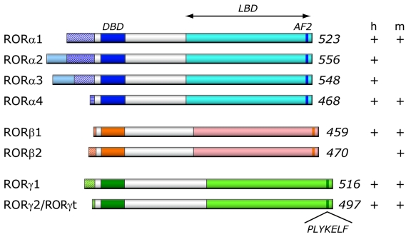
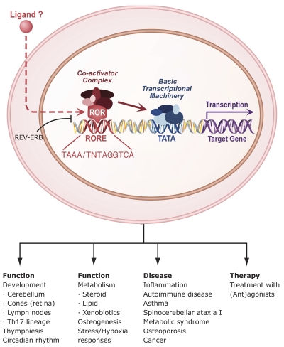
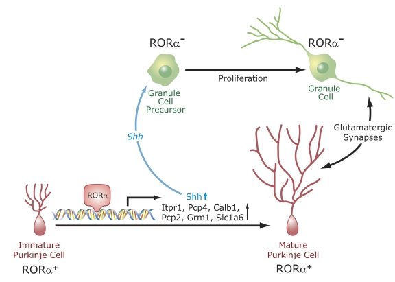
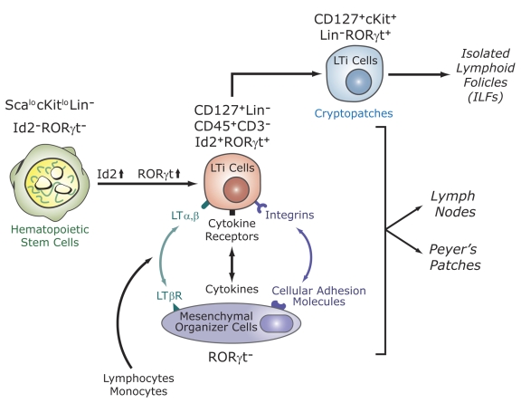
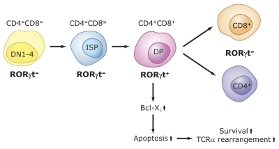
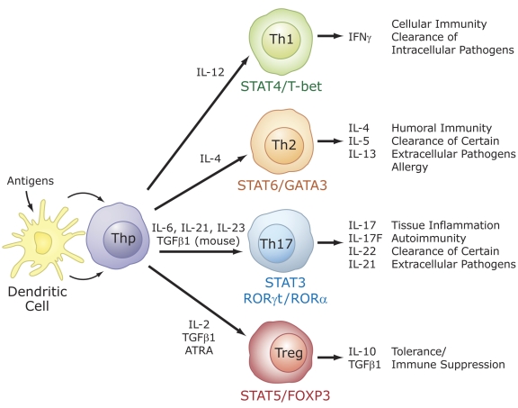
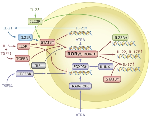
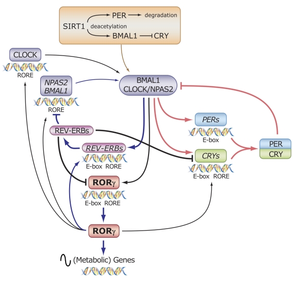
The last few years have witnessed a rapid increase in our knowledge of the retinoid-related orphan receptors RORalpha, -beta, and -gamma (NR1F1-3), their mechanism of action, physiological functions, and their potential role in several pathologies. The characterization of ROR-deficient mice and gene expression profiling in particular have provided great insights into the critical functions of RORs in the regulation of a variety of physiological processes. These studies revealed that RORalpha plays a critical role in the development of the cerebellum, that both RORalpha and RORbeta are required for the maturation of photoreceptors in the retina, and that RORgamma is essential for the development of several secondary lymphoid tissues, including lymph nodes. RORs have been further implicated in the regulation of various metabolic pathways, energy homeostasis, and thymopoiesis. Recent studies identified a critical role for RORgamma in lineage specification of uncommitted CD4+ T helper cells into Th17 cells. In addition, RORs regulate the expression of several components of the circadian clock and may play a role in integrating the circadian clock and the rhythmic pattern of expression of downstream (metabolic) genes. Study of ROR target genes has provided insights into the mechanisms by which RORs control these processes. Moreover, several reports have presented evidence for a potential role of RORs in several pathologies, including osteoporosis, several autoimmune diseases, asthma, cancer, and obesity, and raised the possibility that RORs may serve as potential targets for chemotherapeutic intervention. This prospect was strengthened by recent evidence showing that RORs can function as ligand-dependent transcription factors.


Figures

Figure 1
Figure 1. Schematic representation of ROR family members.
Schematic structure of the various human (h) and mouse (m) ROR isoforms. The DNA binding domain (DBD) and ligand binding domain (LBD), and activation function 2 (AF2) are indicated. The variable regions at the N-terminus of each ROR generated by alternative promoter usage and/or alternative slicing are indicated by patterned boxes on the left. The numbers on the right refer to the total number of amino acids in RORs. The different ROR isoforms identified in human and mouse are shown on the right (+/-).
Figure 2
Figure 2. Mechanism of action of RORs, physiological functions and roles in disease.
RORs bind as a monomer to ROREs consisting of the GGTCA consensus core motif preceded by a 6A/T rich region. REV-ERBs can compete with RORs for binding to ROREs. RORs interact with coactivators or corepressors to positively or negatively regulate gene transcription. RORs are critical in the regulation of many physiological processes and may have a role in several pathologies. Although evidence has been provided indicating that certain ligands can modulate ROR transcriptional activity, whether ROR activity is modulated in vivo by endogenous ligands has yet to be determined. RORs might serve as potential novel targets for chemotherapeutic strategies to intervene in various disease processes.
Figure 3
Figure 3. Regulation of Purkinje cell maturation and cerebellar development by RORα.
RORα regulates the expression of several genes in Purkinje cells. RORα becomes highly expressed in postmitotic Purkinje cells. It regulates their maturation, particularly dendritic differentiation. Dendritogenesis and the expression of several genes, including Shh, Itpr1, Pcp4, Calb1, Pcp2, and Slc1a6, normally expressed in mature Purkinje cells, are inhibited in RORα-deficient mice. The transcription of several of these genes is under direct control of RORα. Shh released by Purkinje cells interacts with Patched (Ptch) receptors on granule cell precursors, leading to activation of GLI transcription factors and the subsequent induction of proliferation-promoting genes. Reduced Shh expression in Purkinje cells from RORα-deficient mice is a major factor in the cerebellar atrophy observed in these mice. Cerebellar granule cells and Purkinje cells mutually interact (e.g., glutamatergic synapses).
Figure 4
Figure 4. RORγt is essential for the development of secondary lymphoid tissues.
Lymphoid tissue inducer (LTi) cells are derived from fetal liver hematopoietic stem cells. This differentiation is accompanied by induction of the transcription factors Id2 and RORγt. LTi cells are recruited from the circulation by mesenchymal organizer cells. This is mediated by multiple receptor-ligand interactions. LTi cells are required for the development of lymph nodes, Peyer’s patches, cryptopatches, and isolated lymphoid follicles (ILFs), which are thought to be derived from cryptopatches after the colonization of the intestine by bacteria. Recently, a novel lymphocyte population (NKp46+NK1.1intCD127+RORγthi) was identified in the gut that may be derived from LTi-like cells in cryptopatches. RORγt is required for the generation and/or survival of LTi cells. The absence of LTi cells in RORγ-deficient mice is responsible for the lack of lymph nodes, Peyer’s patches, cryptopatches, ILFs, and NKp46+NK1.1intCD127+RORγthi cells.
Figure 5
Figure 5. Role of RORγt in thymopoiesis.
CD4-CD8-CD25-CD44+ (DN1) cells differentiate via DN2, DN3, DN4 into immature single positive (ISP) cells (CD3-CD4-CD8low). These cells subsequently differentiate into CD3+CD4+CD8+, DP thymocytes. RORγt, as well as Bcl-XL, are induced during the ISP-DP transition and again down-regulated during the differentiation of DP into CD4+ and CD8+ single positive cells. RORγt promotes the differentiation of ISP into DP cells and positively regulates the expression of the anti-apoptotic gene Bcl-XL. The latter enhances cell survival that subsequently promotes TCR rearrangements. Lack of RORγt expression inhibits the ISP-DP transition and reduces expression of Bcl-XL in DP thymocytes. The latter results in accelerated apoptosis, reduced lifespan of DP thymocytes and, consequently, impaired TCRα rearrangements
Figure 6
Figure 6. Specific role for RORs in T cell lineage specification.
Differentiation into different effector CD4+ T cell lineages, T helper (Th) 1, Th2, Th17, and T regulatory (Treg) cells is initiated through an interaction of dendritic cells with uncommitted (naïve) CD4+ T helper cells (Thp). The effector cell types are characterized by their synthesis of specific cytokines and their immuno-regulatory functions, as indicated on the right. The differentiation along different lineages involves different cytokines and the activation of distinct signaling cascades and transcription factors that result in the induction of additional cyto/chemokines and cyto/chemokine receptors, which may be part of positive and negative feedback loops. These cytokines and transcription factors may favor one cell lineage, while inhibiting another (not indicated). RORα and RORγt are induced during differentiation of Thp into the Th17 lineage and are critical for this lineage specification.
Figure 7
Figure 7. RORs are essential for Th17 cell differentiation and IL-17 expression.
In the presence of TCR engagement, treatment of Thp cells with IL-6 plus TGFβ1 induces differentiation along the Th17 lineage and the activation of several genes, including IL-21, RORα, RORγt, IL-17, IL-17F, IL-22, and IL23R. Interaction of IL-23 and IL-21 with, respectively, IL23R and IL21R, reinforce Th17 differentiation and ROR expression. RORα and RORγt are required for the induction of IL-17, IL-17F, and IL23R, but not IL-21. A balance between FOXP3, which is induced by treatment with TGFβ alone, determines whether Thp cells differentiate into Th17 or Treg cells. ATRA, through activation of RARα-RXR complexes, leads to increased FOXP3 and reduced ROR expression. FOXP3 inhibits RORα and RORγt transcriptional activity by interacting directly with RORs, thereby promoting differentiation along the Treg lineage and inhibiting Th17 differentiation. Transcriptional regulators IRF4 and RUNX1 have also been implicated in the regulation of Treg and Th17 differentiation.
Figure 8
Figure 8. RORs function as integrators of circadian oscillators and the rhythmic expression of downstream (metabolic) genes.
The rhythmic expression of RORs is greatly dependent on the ROR isoform and tissue type. The core loop of the oscillator consists of the heterodimeric activators BMAL1 and CLOCK that activate the expression of PERs and CRYs. PER and CRY heterodimers interact with the BMAL1-CLOCK complex and negatively regulate their transcriptional activity. BMAL1 and CLOCK also activate the expression of REV-ERBs, which through their interaction with ROREs repress the transcription of several genes, including BMAL1, CRY1, and RORγ. REV-ERBs are critical in the rhythmic expression of BMAL1, CRY1 and RORγ in liver and several other tissues. RORγ does not affect the rhythmic expression, but increases the peak level of expression of BMAL1, CRY1, NPAS2, CLOCK, and REV-ERBα. The major function of RORγ appears to be to regulate the oscillatory pattern of expression of downstream target genes and, therefore, the rhythmic nature of several physiological processes. RORα exhibits a very weak rhythmic pattern of expression in liver and its major function also appears to be in regulating the (rhythmic) expression of downstream target genes. SIRT, through its deacetylase activity, promotes the degradation of PER and inhibits the interaction of CRY with BMAL1/CLOCK complex, thereby enhancing BMAL1/CLOCK transcriptional activity.

Similar articles

Cited by

References

    1. Acosta-Rodriguez E. V., Napolitani G., Lanzavecchia A., Sallusto F. Interleukins 1beta and 6 but not transforming growth factor-β are essential for the differentiation of interleukin 17-producing human T helper cells. Nat Immunol. 2007;8:942–9. - PubMed
    1. Akashi M., Takumi T. The orphan nuclear receptor RORalpha regulates circadian transcription of the mammalian core-clock Bmal1. Nat Struct Mol Biol. 2005;12:441–8. - PubMed
    1. Akimzhanov A. M., Yang X. O., Dong C. Chromatin remodeling of interleukin-17 (IL-17)-IL-17F cytokine gene locus during inflammatory helper T cell differentiation. J Biol Chem. 2007;282:5969–72. - PubMed
    1. Akiyama T. E., Gonzalez F. J. Regulation of P450 genes by liver-enriched transcription factors and nuclear receptors. Biochim Biophys Acta. 2003;1619:223–34. - PubMed
    1. Albrecht U. Invited review: regulation of mammalian circadian clock genes. J Appl Physiol. 2002;92:1348–55. - PubMed

Publication types

MeSH terms

Substances