Review
. 2009 Sep;94(3):299-312.
doi: 10.1007/s11060-009-9875-7. Epub 2009 Apr 21.

Rat brain tumor models in experimental neuro-oncology: the C6, 9L, T9, RG2, F98, BT4C, RT-2 and CNS-1 gliomas

Affiliations
Review

Rat brain tumor models in experimental neuro-oncology: the C6, 9L, T9, RG2, F98, BT4C, RT-2 and CNS-1 gliomas

Rolf F Barth et al. J Neurooncol. 2009 Sep.

Abstract

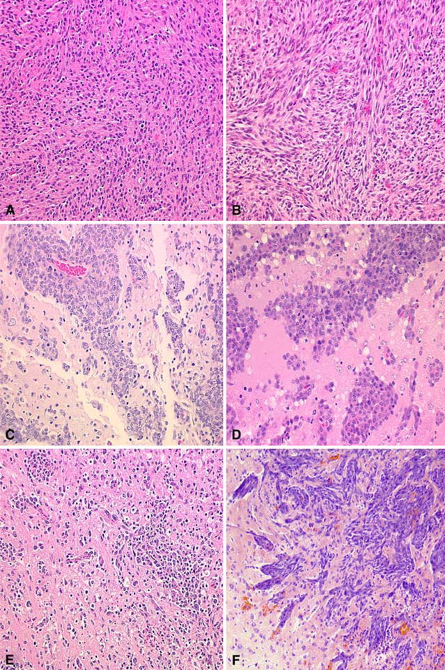
In this review we will describe eight commonly used rat brain tumor models and their application for the development of novel therapeutic and diagnostic modalities. The C6, 9L and T9 gliomas were induced by repeated injections of methylnitrosourea (MNU) to adult rats. The C6 glioma has been used extensively for a variety of studies, but since it arose in an outbred Wistar rat, it is not syngeneic to any inbred strain, and its potential to evoke an alloimmune response is a serious limitation. The 9L gliosarcoma has been used widely and has provided important information relating to brain tumor biology and therapy. The T9 glioma, although not generally recognized, was and probably still is the same as the 9L. Both of these tumors arose in Fischer rats and can be immunogenic in syngeneic hosts, a fact that must be taken into consideration when used in therapy studies, especially if survival is the endpoint. The RG2 and F98 gliomas were both chemically induced by administering ethylnitrosourea (ENU) to pregnant rats, the progeny of which developed brain tumors that subsequently were propagated in vitro and cloned. They are either weakly or non-immunogenic and have an invasive pattern of growth and uniform lethality, which make them particularly attractive models to test new therapeutic modalities. The CNS-1 glioma was induced by administering MNU to a Lewis rat. It has an infiltrative pattern of growth and is weakly immunogenic, which should make it useful in experimental neuro-oncology. Finally, the BT4C glioma was induced by administering ENU to a BD IX rat, following which brain cells were propagated in vitro until a tumorigenic clone was isolated. This tumor has been used for a variety of studies to evaluate new therapeutic modalities. The Avian Sarcoma Virus (ASV) induced tumors, and a continuous cell line derived from one of them designated RT-2, have been useful for studies in which de novo tumor induction is an important requirement. These tumors also are immunogenic and this limits their usefulness for therapy studies. It is essential to recognize the limitations of each of the models that have been described, and depending upon the nature of the study to be conducted, it is important that the appropriate model be selected.


Figures

Fig. 1
Fig. 1
Histopathologic features of the C6, 9L, RG2, F98, CNS-1, and BT4C brain tumors. A The C6 glioma is composed of a pleomorphic population of cells with nuclei ranging from round to oblong. A herring-bone pattern of growth is seen in some areas and there is focal invasion of contiguous normal brain. There are scattered foci of necrosis with pseudo-palisading of tumor cells at the periphery. B The 9L gliosarcoma is composed of spindle-shaped cells with a sarcomatoid appearance. A whorled pattern of growth is seen with sharp delineation of the margins of the tumor with little invasion of contiguous normal brain. C The RG2 glioma is very similar in appearance to the F98 glioma and also has a highly invasive pattern of growth. D The F98 glioma is composed of a mixed population of spindle-shaped cells with fusiform nuclei, frequently forming a whorled pattern of growth, and a smaller subpopulation of polygonal cells with round to oval nuclei. There is extensive invasion of contiguous normal brain with islands of tumor cells at varying distances from the main tumor mass, which form perivascular clusters. Usually, there is a central area of necrosis filled with tumor cell ghosts. E The CNS-1 glioma is composed of a pleomorphic population of cells that show great variation in size and shape. There is extensive invasion of contiguous normal brain with dense infiltrates in some areas and in others, more circumscribed clusters of tumor cells. Small foci of hemorrhage are scattered through the tumor. F The BT4C glioma is composed of a pleomorphic population of tumor cells with a sarcomatous pattern of growth. Scattered tumor giant cells are seen and mitotic figures are frequent. The tumor grows expansively and invades the surrounding normal brain along perivascular tracts and occasional tumor cell nests are seen in the surrounding normal brain. There is neo-vascularization, especially in the tumor periphery, where microhemorrhages are frequent. Central necrosis is usually not present but occasionally scattered areas of necrosis may be seen in larger tumors. (Photomicrograph of the BT4C was kindly provided by M. Sandström and description by M. Johansson. Representative microscope slides of the CNS-1 glioma were kindly provided by Dr. Carol Kruse). All photomicrographs are at a magnification of 200×, except for F

References

    1. Fomchenko EI, Holland EC. Mouse models of brain tumors and their applications in preclinical trials. Clin Cancer Res. 2006;12:5288–5297. doi: 10.1158/1078-0432.CCR-06-0438. - DOI - PubMed
    1. Lampson LA. New animal models to probe brain tumor biology, therapy, and immunotherapy: advantages and remaining concerns. J Neurooncol. 2001;53:275–287. doi: 10.1023/A:1012230113527. - DOI - PubMed
    1. Kimmelman J, Nalbantoglu J. Faithful companions: a proposal for neurooncology trials in pet dogs. Cancer Res. 2007;67:4541–4544. doi: 10.1158/0008-5472.CAN-06-3792. - DOI - PubMed
    1. Krushelnycky BW, Farr-Jones MA, Mielke B, et al. Development of a large-animal human brain tumor xenograft model in immunosuppressed cats. Cancer Res. 1991;51:2430–2437. - PubMed
    1. Barth RF. Rat brain tumor models in experimental neuro-oncology: the 9L, C6, T9, F98, RG2 (D74), RT-2 and CNS-1 gliomas. J Neurooncol. 1998;36:91–102. doi: 10.1023/A:1005805203044. - DOI - PubMed