

Local expression of interferon-alpha and interferon receptors in cervical intraepithelial neoplasia
- PMID: 19381629
- PMCID: PMC11030549
- DOI: 10.1007/s00262-009-0707-6
Local expression of interferon-alpha and interferon receptors in cervical intraepithelial neoplasia
Abstract
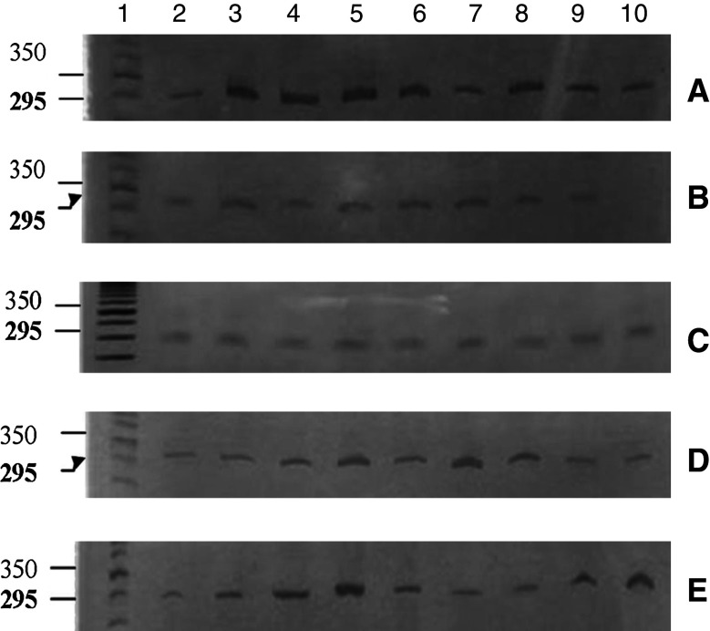
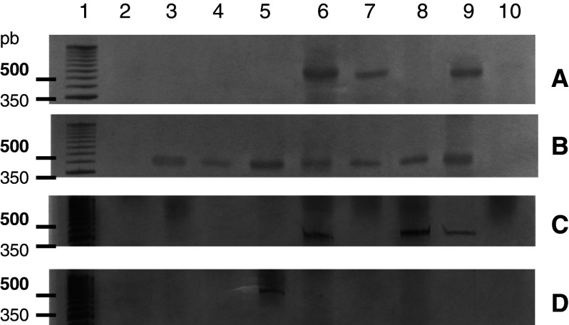
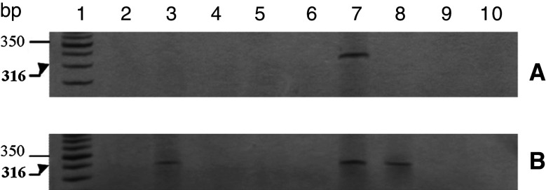
Purpose: The present study evaluated mRNA expression of interferon-alpha (IFN-alpha), IFN-alpha receptor subunits (IFNAR-1 and IFNAR-2) and an IFN-stimulated gene encoding the enzyme 2',5'-oligoadenylate synthetase (2'5'OAS) in biopsies on patients with varying grades of cervical intraepithelial neoplasia (CIN I, II and III).
Methods: Uterine cervix biopsies were collected from women with CIN I, II and III (n = 28) and controls without CIN lesions or human papilloma virus (HPV) infection (n = 17). The presence of high and low-risk HPV DNA was determined using hybrid capture. The mRNA levels of IFNAR-1, IFNAR-2, IFN-alpha and 2'5'OAS were determined by RT-PCR with specific primers.
Results: The control group exhibited a greater frequency of IFNAR-1 expression (10/17; 58.3%) than the CIN samples (4/28; 14.2%) (P = 0.0018), while, the expression of IFNAR-2 was also greater in the control samples (11/17; 64.7%) than in the patients with lesions (2/28; 7.1%) (P = 0.0018). Importantly, simultaneous expression of both receptors was observed only in the control group (8/17; 47.0%) (P = 0.0001). Among the CIN samples, there was one case of low expression of mRNA of IFNAR-1 and IFNAR-2. IFN-alpha was present in 14.2% (4/28) of the CIN samples but was not expressed in the control group. mRNA 2'5'OAS were expressed in 28.5% (8/28) of the CIN samples and 11.7% (2/17) of the control samples (not statistically significant). Fifty percent (14/28) of the CIN samples were positive for HPV DNA.
Conclusions: Cervical biopsy samples from control women or those without neoplasia or HPV infection displayed higher IFN-alpha receptor expression than those with CIN, while simultaneous expression of both IFN-alpha receptor subunits was found only in the control group. There was no significant difference in mRNA expression of IFN-alpha and 2'5'OAS between the control and CIN groups. Then we concluded that the samples obtained from patients with CIN present low levels of the IFN-alpha receptor mRNA.
Figures


Similar articles
-
Increased expression of programmed death (PD)-1 and its ligand PD-L1 correlates with impaired cell-mediated immunity in high-risk human papillomavirus-related cervical intraepithelial neoplasia.Immunology. 2013 Aug;139(4):513-22. doi: 10.1111/imm.12101. Immunology. 2013. PMID: 23521696 Free PMC article.
-
Physical state and expression of HPV DNA in benign and dysplastic cervical tissue: different levels of viral integration are correlated with lesion grade.Gynecol Oncol. 2004 Mar;92(3):873-80. doi: 10.1016/j.ygyno.2003.11.035. Gynecol Oncol. 2004. PMID: 14984955
-
Cytokine production patterns in cervical intraepithelial neoplasia: association with human papillomavirus infection.J Natl Cancer Inst. 1997 Feb 5;89(3):245-50. doi: 10.1093/jnci/89.3.245. J Natl Cancer Inst. 1997. PMID: 9017005
-
Accumulation of invariant NKT cells with increased IFN-γ production in persistent high-risk HPV-infected high-grade cervical intraepithelial neoplasia.Diagn Pathol. 2015 Apr 2;10:20. doi: 10.1186/s13000-015-0254-8. Diagn Pathol. 2015. PMID: 25885042 Free PMC article.
-
The importance of alpha/beta (alpha/13) interferon receptors and signaling pathways for the treatment of cervical intraepithelial neoplasias.Eur J Gynaecol Oncol. 2014;35(4):368-72. Eur J Gynaecol Oncol. 2014. PMID: 25118475 Review.
Cited by
-
HPV Induces Changes in Innate Immune and Adhesion Molecule Markers in Cervical Mucosa With Potential Impact on HIV Infection.Front Immunol. 2020 Sep 3;11:2078. doi: 10.3389/fimmu.2020.02078. eCollection 2020. Front Immunol. 2020. PMID: 33013878 Free PMC article.
-
The human papillomavirus E7 oncoprotein as a regulator of transcription.Virus Res. 2017 Mar 2;231:56-75. doi: 10.1016/j.virusres.2016.10.017. Epub 2016 Nov 8. Virus Res. 2017. PMID: 27818212 Free PMC article. Review.
-
The Interaction Between Human Papillomaviruses and the Stromal Microenvironment.Prog Mol Biol Transl Sci. 2016;144:169-238. doi: 10.1016/bs.pmbts.2016.09.003. Epub 2016 Oct 11. Prog Mol Biol Transl Sci. 2016. PMID: 27865458 Free PMC article. Review.
-
Pathways of IFN-alpha Activation in Patients with Cervical Intraepithelial Neoplasia (CIN).Rev Bras Ginecol Obstet. 2021 Sep;43(9):682-689. doi: 10.1055/s-0041-1735301. Epub 2021 Oct 20. Rev Bras Ginecol Obstet. 2021. PMID: 34670303 Free PMC article.
References
-
- Arany I, Goel A, Tyring SK. Interferon response depends on viral transcription in human papillomavirus-containing lesions. Anticancer Res. 1995;15:2865–2869. - PubMed
-
- Chieux V, Hober D, Chehadeh W, Wattr P. Anti-viral proteins: from interferon alpha to its receptor. Ann Biol Clin. 1999;57(3):283–290. - PubMed
-
- Cintorino M, Tripodi SA, Romagnoli R, Ietta F, Ricci MG, Paulesu L. Interferons and their receptors in human papillomavirus lesions of the uterine cervix. Eur J Gynaecol Oncol. 2002;23:145–150. - PubMed
Publication types
MeSH terms
Substances
LinkOut - more resources
Full Text Sources
Other Literature Sources
Medical
