

doi: 10.1007/978-1-59745-190-1_5.
Visualization of the expression of HMGN nucleosomal binding proteins in the developing mouse embryo and in adult mouse tissues
Affiliations
- PMID: 19381921
- PMCID: PMC3402339
- DOI: 10.1007/978-1-59745-190-1_5
Item in Clipboard
Visualization of the expression of HMGN nucleosomal binding proteins in the developing mouse embryo and in adult mouse tissues
Methods Mol Biol.
2009.
Abstract
Visualization of the expression pattern of specific proteins during development and in adult tissues provides important clues as to their possible role in various cellular processes. Mouse is the organism of choice for obtaining information on gene expression patterns in higher eukaryotes. This chapter describes the protocols we utilized to visualize Hmgn transcripts and HMGN proteins in mouse tissues. HMGN are chromatin-binding proteins that affect chromatin structure and function and play a role in cellular differentiation.
Figures
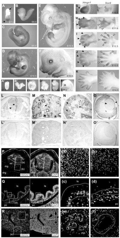
Expression patterns of Hmgn1, Sox9, and HMGN1 protein in the developing mouse embryo. (A–E) Whole mount in situ hybridization using mouse Hmgn1 antisense probe. Pictures of A–C are taken with microscopy and D–E are taken with digital camera that makes color appearance different (both AP staining, same substrate NBT-BCIP is used). (A) At E7.5 embryo, Hmgn1 is detected in the embryonic region (arrowhead) while no signal is detected in the ectoplacental cone (arrow). (A–F) As the embryo develops, the signal is detected only in the surface because of difficulty of probe penetration. (A′–F′) Control experiments using mouse Hmgn1 sense probe. (G–K) The comparison of expression patterns of Hmgn1 (left) and Sox9 (right) in the developing limb bud. Hmgn1 expression is detected in the distal mesenchyme (filled arrowhead in G; left) and interdigit mesenchyme (filled arrowheads in H–J; left), while Sox9 is detected in the compensating mesenchyme (open arrow head in G; right) and digit mesenchyme (open arrowheads H–J; right). (L–O) Expression patterns of Hmgn1 in the histological sections of E14.5 embryo detected by in situ hybridization. (L) Section through neural tube. Hmgn1 is strongly expressed in the ventricular zone (filled arrow head), surface ectoderm (blank arrow head), and weakly expressed in the mantle layer (arrow). (M) Section through lung. Hmng1 is strongly expressed in the distal epithelium (arrow head), while no expression is detected in the bronchiole (arrow). (N) Section through kidney. Hmgn1 is expressed interstitial mesenchymal cells around the urinary tubules (arrow head) and glomerulus (arrow). Expression level is stronger in the peripheral than in the central region of the kidney. (O) Section through eye (1). Strong expression of Hmgn1 is observed in the retina (arrow head) and lens epithelium, but not in the lens fiber (open arrow head). (L′–O′) Control experiments using mouse Hmgn1 sense probe. No significant signals are detected. (P–R) The localization of HMGN1 protein is detected by immunofluorescence. Cell nuclei are counter stained with DAPI. (P, a and b) Sections through the E14.5 neural tube. HMGN1 is localized to the roof plate (rp), ependymal layer (el) of the neural tube, dorsal region of mantle layer (mL and (a) in P) but downregulated in the dorsal region of mantle layer (b in P) and absent from marginal layer. mg, marginal layer; dg, dorsal root ganglia. (Q, c, and d) Sections through the developing mouse stomach at E16.5. HMGN1 expression is localized to basal layer of gastric mucosa (bl in (c)) and longitudinal muscle layer (lm in (d)). ls, lumen of stomach; pc, peritoneal cavity; lv, liver; cm, circular muscle layer. (R, e, and f) Sections through the lung. At E16.5, bronchial expression of HMGN1 was downregulated in the proximal region (br in (f)) and decreased in the mesenchymal region but remained strong in the distal region (de, distal epithelium in (e)); me, mesenchyme.
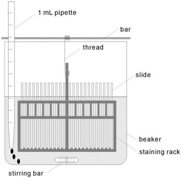
Acetylation buffer treatment. This figure illustrates how to add 1 mL of acetic anhydrate little by little. For detail, see
Note 14.
Similar articles
-
Developmental function of HMGN proteins.Biochim Biophys Acta. 2010 Jan-Feb;1799(1-2):69-73. doi: 10.1016/j.bbagrm.2009.11.011. Biochim Biophys Acta. 2010. PMID: 20123069 Free PMC article. Review.
-
HMGN5/NSBP1: a new member of the HMGN protein family that affects chromatin structure and function.Biochim Biophys Acta. 2010 Jan-Feb;1799(1-2):86-92. doi: 10.1016/j.bbagrm.2009.09.012. Biochim Biophys Acta. 2010. PMID: 20123071 Free PMC article. Review.
-
Developmental role of HMGN proteins in Xenopus laevis.Mech Dev. 2003 Oct;120(10):1177-92. doi: 10.1016/j.mod.2003.07.001. Mech Dev. 2003. PMID: 14568106
-
High mobility group N proteins modulate the fidelity of the cellular transcriptional profile in a tissue- and variant-specific manner.J Biol Chem. 2013 Jun 7;288(23):16690-16703. doi: 10.1074/jbc.M113.463315. Epub 2013 Apr 24. J Biol Chem. 2013. PMID: 23620591 Free PMC article.
-
Epigenetic Regulation of Ameloblast Differentiation by HMGN Proteins.J Dent Res. 2024 Jan;103(1):51-61. doi: 10.1177/00220345231202468. Epub 2023 Nov 10. J Dent Res. 2024. PMID: 37950483 Free PMC article.
References
-
- Bustin M. Chromatin unfolding and activation by HMGN(*) chromosomal proteins. Trends Biochem Sci. 2001;26:431–7. - PubMed
Publication types
MeSH terms
Substances
Grants and funding
LinkOut - more resources
Full Text Sources
