. 2009 May;13(5):866-86.
doi: 10.1111/j.1582-4934.2009.00758.x. Epub 2009 Apr 20.

Cardiac renewing: interstitial Cajal-like cells nurse cardiomyocyte progenitors in epicardial stem cell niches

Affiliations

Cardiac renewing: interstitial Cajal-like cells nurse cardiomyocyte progenitors in epicardial stem cell niches

L M Popescu et al. J Cell Mol Med. 2009 May.

Abstract

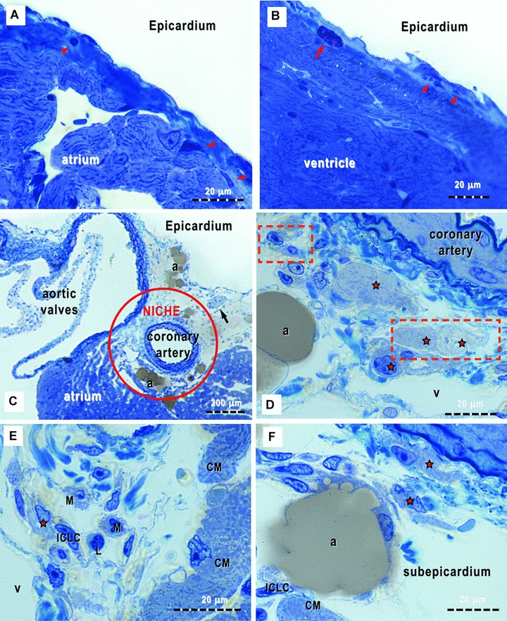
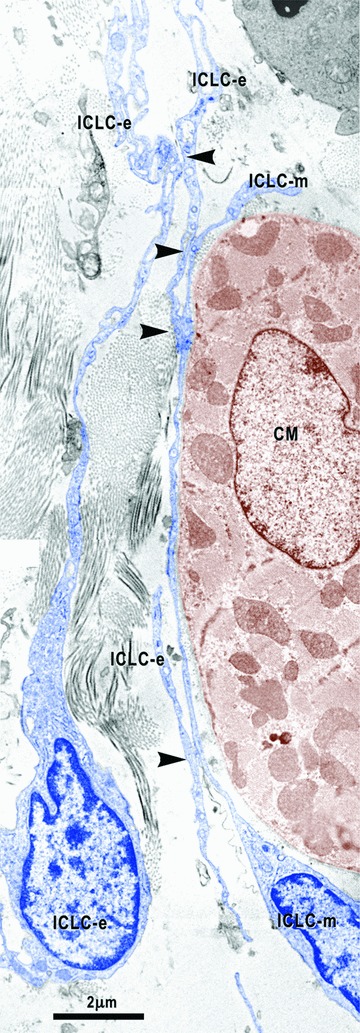
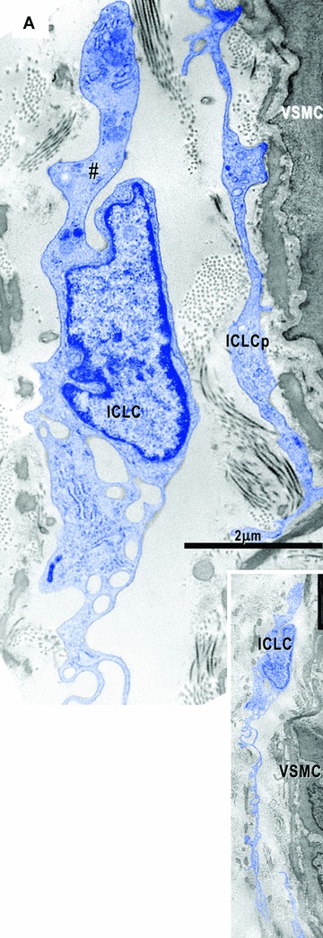
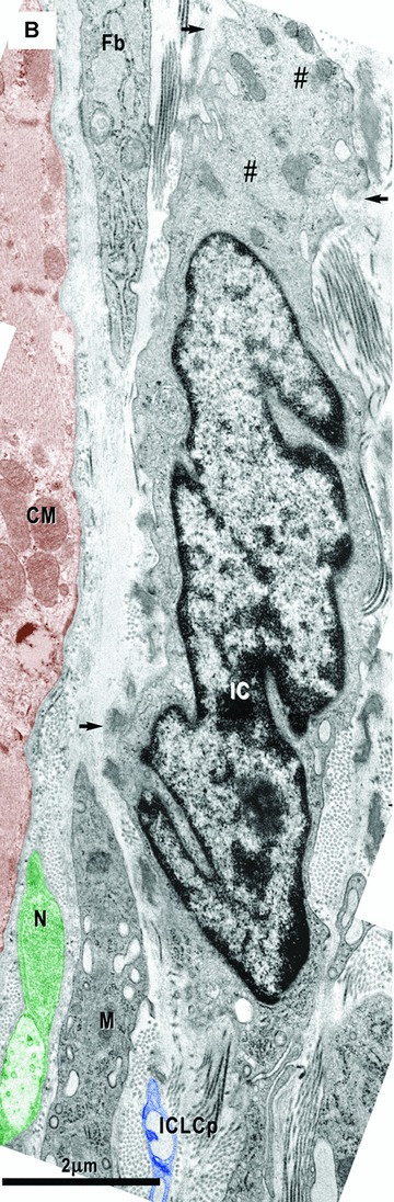
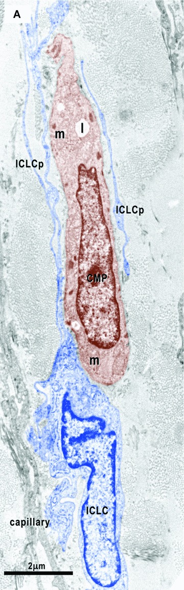
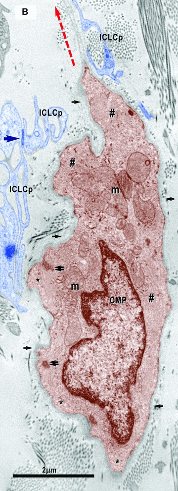
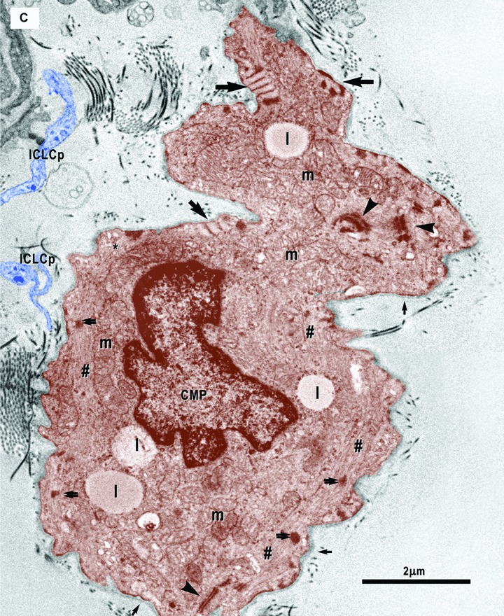
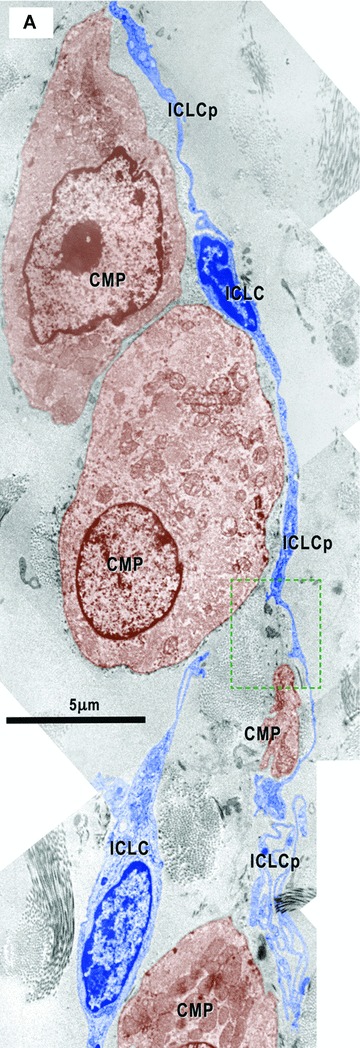
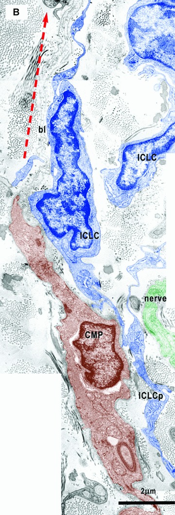
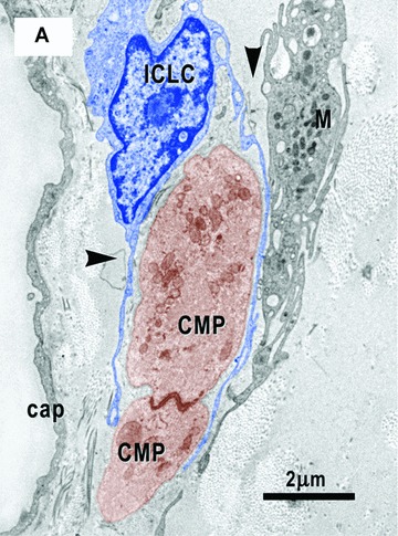
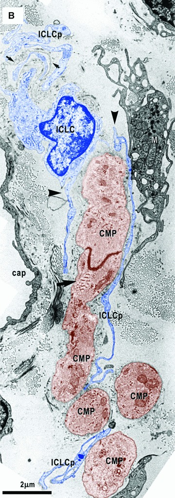
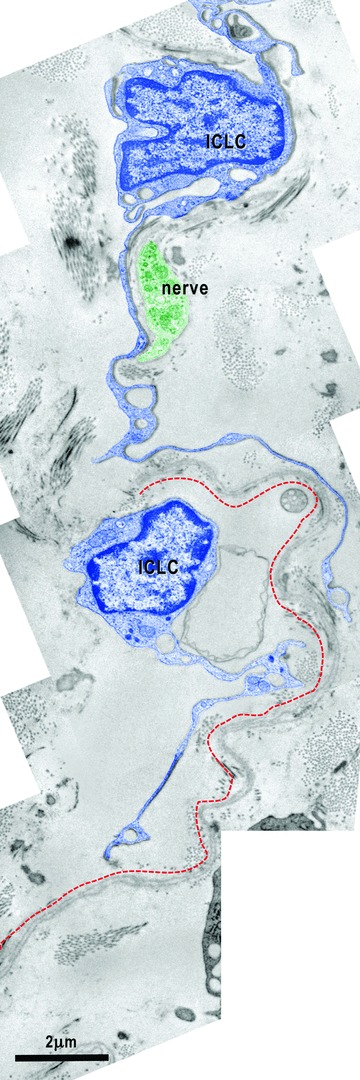
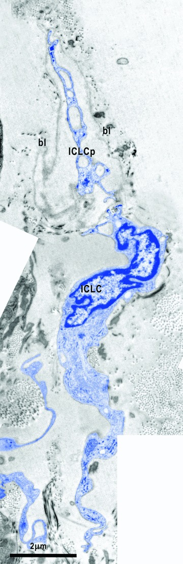
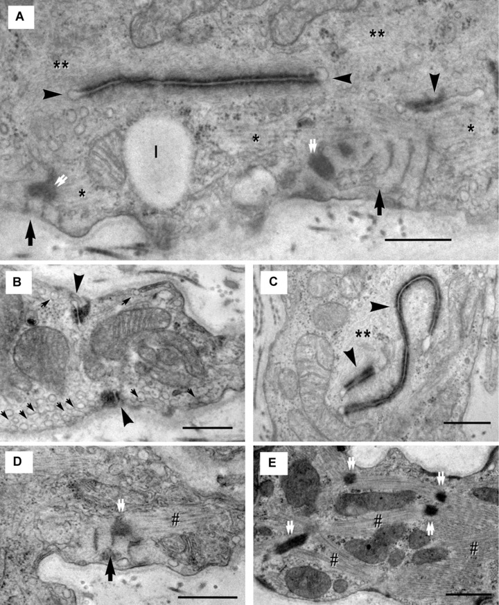
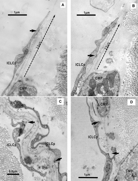
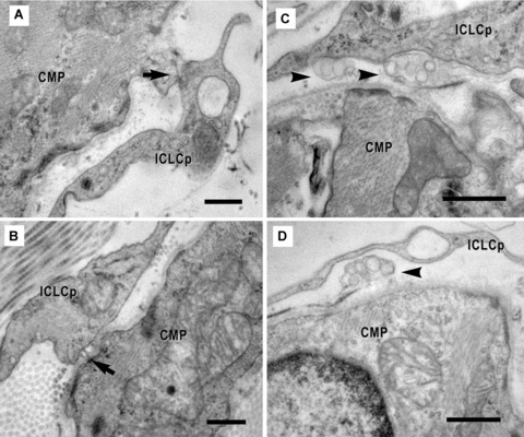
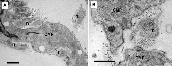
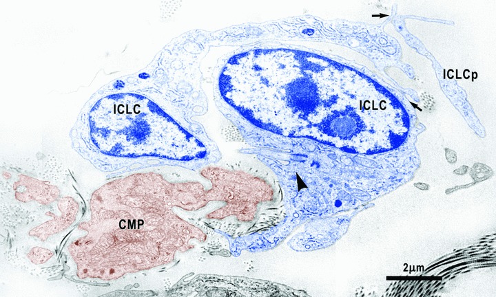
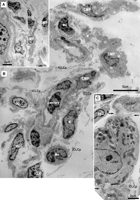
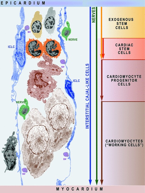
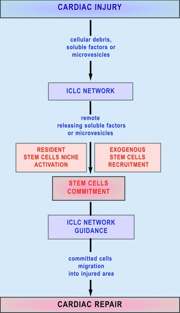
Recent studies suggested that various cell lineages exist within the subepicardium and we supposed that this area could host cardiac stem cell niches (CSCNs). Using transmission electron microscopy, we have found at least 10 types of cells coexisting in the subepicardium of normal adult mice: adipocytes, fibroblasts, Schwann cells and nerve fibres, isolated smooth muscle cells, mast cells, macrophages, lymphocytes, interstitial Cajal-like cells (ICLCs) and cardiomyocytes progenitors (CMPs). The latter cells, sited in the area of origin of coronary arteries and aorta, showed typical features of either very immature or developing cardiomyocytes. Some of these cells were connected to each other to form columns surrounded by a basal lamina and embedded in a cellular network made by ICLCs. Complex intercellular communication occurs between the ICLCs and CMPs through electron-dense nanostructures or through shed vesicles. We provide here for the first time the ultrastructural description of CSCN in the adult mice myocardium, mainly containing ICLCs and CMPs. The existence of resident CMPs in different developmental stages proves that cardiac renewing is a continuous process. We suggest that ICLCs might act as supporting nurse cells of the cardiac niches and may be responsible for activation, commitment and migration of the stem cells out of the niches. Briefly, not only resident cardiac stem cells but also ICLCs regulate myocyte turnover and contribute to both cardiac cellular homeostasis and endogenous repair/remodelling after injuries.


Figures

1
1
(A)-(F). Light microscopy of toluidine-blue stained semi-thin sections (∼1 μm, or less) of Epon-embedded epicardium from 16-week-old mice (supposed to be the equivalent of 25 human years). The loose connective tissue with very few cells (arrowheads) may be observed immediately below the epicardial mesothelium (the so-called subepicardium), in both cases of atrium (A) and ventricle (B). A metachromatically stained mast cell (red arrow) can be easily recognized. (C) shows the richness of cell population types surrounding the coronary artery and a cross sectioned nerve (arrow). Red circle marks the epicardial stem cell niche. (D)-(F) demonstrate different cellular silhouettes scattered in the loose connective tissue of the cardiac stem cell niche. Note, the presence of ‘young’ cardiomyocytes (red stars); the orange rectangles areas correspond to high-resolution electron microscope images in Fig. 6. ICLC – interstitial Cajal-like cells; CM – cardiomyocytes; L – lymphocyte; M – macrophage; v – venules and a – adipocytes.
2
2
Digitally coloured image shows the close relationships between the epicardial ICLC (ICLC-e) and the ICLC bordering the myocardium (ICLC-m). Arrowheads indicate close contacts between epicardial ICLC and myocardial ICLC. ICLC – interstitial Cajal-like cells are blue coloured and CM – cardiomyocyte coloured in brown.
3
3
Electron micrographs show interstitial cells in the subepicardial area next to the coronary artery. (A) – Interstitial Cajal-like cells (ICLC, blue) have long and thin processes (inset), a lacunar cytoplasm, few cisternae of the rough endoplasmic reticulum and intermediate filaments (#). No basal lamina appears surrounding these cells. Another ICLC process (ICLCp) is near coronary smooth muscle cells (VSMC). (B) – Next to the atrial myocardium, an interstitial cell (IC) particularly rich in filaments (#) shows focal basal lamina-like material (arrows). N – nerve (green); M – macrophage; Fb – fibroblast; CM – cardiomyocyte (brown).
3
3
Electron micrographs show interstitial cells in the subepicardial area next to the coronary artery. (A) – Interstitial Cajal-like cells (ICLC, blue) have long and thin processes (inset), a lacunar cytoplasm, few cisternae of the rough endoplasmic reticulum and intermediate filaments (#). No basal lamina appears surrounding these cells. Another ICLC process (ICLCp) is near coronary smooth muscle cells (VSMC). (B) – Next to the atrial myocardium, an interstitial cell (IC) particularly rich in filaments (#) shows focal basal lamina-like material (arrows). N – nerve (green); M – macrophage; Fb – fibroblast; CM – cardiomyocyte (brown).
4
4
(A)–(C). Digitally coloured electron micrographs show cardiomyocyte progenitors (CMP, brown) in different stages of development and ICLC (blue). CMP have features of immature cardiomyocytes: a high nucleo-cytoplasmatic ratio and a continuous basal lamina (small arrows), numerous mitochondria (m) and ribosomes, few endoplasmic reticulum cisternae, some lipid droplets (l) and caveolae (*). These cells contain unorganized bundles of filaments (#) attached to electron dense structures (double arrows) similar to Z bands. (A) – ICLC processes (ICLCp) closely accompany the immature cardiomyocyte. B– The basal lamina exceeds the cellular profile and delimitates a slender acellular space. An ICLC process seems to support and direct this lamina (red dotted arrow). The large arrow indicates a gap junction in between two ICLCp. (C) – Note the presence of the subplasmalemmal leptofibrils (large arrows) and intracellular desmosome-like structures (arrowheads).
4
4
(A)–(C). Digitally coloured electron micrographs show cardiomyocyte progenitors (CMP, brown) in different stages of development and ICLC (blue). CMP have features of immature cardiomyocytes: a high nucleo-cytoplasmatic ratio and a continuous basal lamina (small arrows), numerous mitochondria (m) and ribosomes, few endoplasmic reticulum cisternae, some lipid droplets (l) and caveolae (*). These cells contain unorganized bundles of filaments (#) attached to electron dense structures (double arrows) similar to Z bands. (A) – ICLC processes (ICLCp) closely accompany the immature cardiomyocyte. B– The basal lamina exceeds the cellular profile and delimitates a slender acellular space. An ICLC process seems to support and direct this lamina (red dotted arrow). The large arrow indicates a gap junction in between two ICLCp. (C) – Note the presence of the subplasmalemmal leptofibrils (large arrows) and intracellular desmosome-like structures (arrowheads).
6
6
(A) and (B) Electron micrographs (from the areas marked in Fig. 1D) illustrate the relationships of the ICLC (blue coloured) with cardiomyocyte progenitors (CMP, brown). The ICLC processes (ICLCp) run parallel with the main axis of the CMP and seem to establish their direction of development. In (B) it is clear that the ICLC sustain a direction (red dotted arrow) for the basal lamina (bl) which goes beyond the CMP. Details from the upper area in Fig. 10C. The square mark area from A is enlarged in Fig. 10D.
6
6
(A) and (B) Electron micrographs (from the areas marked in Fig. 1D) illustrate the relationships of the ICLC (blue coloured) with cardiomyocyte progenitors (CMP, brown). The ICLC processes (ICLCp) run parallel with the main axis of the CMP and seem to establish their direction of development. In (B) it is clear that the ICLC sustain a direction (red dotted arrow) for the basal lamina (bl) which goes beyond the CMP. Details from the upper area in Fig. 10C. The square mark area from A is enlarged in Fig. 10D.
7
7
(A) and (B) Electron micrographs on serial sections show an ICLC (blue coloured) with two processes (arrowheads) which hold two cardiomyocyte progenitors (CMP, brown). Note close contacts of one ICLC process (ICLCp) with a macrophage (M). (B) – A basal lamina – like material surrounds an ICLC process (small arrows in the upper left corner). Leptomeric fibrils (arrow) connect the primitive intercalated disk with the plasma membrane. Cap – capillary.
7
7
(A) and (B) Electron micrographs on serial sections show an ICLC (blue coloured) with two processes (arrowheads) which hold two cardiomyocyte progenitors (CMP, brown). Note close contacts of one ICLC process (ICLCp) with a macrophage (M). (B) – A basal lamina – like material surrounds an ICLC process (small arrows in the upper left corner). Leptomeric fibrils (arrow) connect the primitive intercalated disk with the plasma membrane. Cap – capillary.
8
8
Electron micrograph illustrates the relationships of the ICLC (blue coloured) with a nerve fibre (green) and the basal lamina material that extends over the bodies of the cardiomyocyte progenitors. The processes of two ICLC guide a 30-μm-long bi-layered basal lamina (red dotted line).
9
9
Electron micrograph illustrates the relationships of one ICLC (blue) with the basal lamina (bl). In this micrograph, one ICLC process (ICLCp) with a lacunar cytoplasm seems to be embedded in the space delimited by a continuous basal lamina.
5
5
(A)-(E). Electron micrographs show ultrastructural features of the cardiomyocyte progenitors. (A) – Desmosome-like structures (arrowheads) are entirely located inside the cell and seem to organize thin filaments (**). Thick filaments (*) appear to be organized by leptofibrils (arrows). Z-like dense structures (double arrows) are visible at the end of leptofibrils. Image in (B) shows numerous caveolae (small arrows) and confluent intracytoplasmatic vesicles (primordial T tubules) and suggests that the desmosome-like structures (arrowheads) start from periphery and extend towards the centre of the cell. In (C), it is obvious that thin filaments (**) are organized by desmosome-like structures (arrowheads). (D) – Myofibrils (#) appear to be directed by leptofibrils (arrow) that anchor them to the plasma membrane throughout Z-like dense structures (double arrow). In (E), the nascent sarcomeres are visible: Z-like dense structures (double arrows) organize myofibrils (#) in an immature sarcomeric configuration. l – lipid droplet. Scale bar = 0.5 μm.
10
10
(A) and (B). Electron micrographs on serial ultrathin sections highlight the connections of the ICLC processes (ICLCp) with the basal lamina of the CMP by electron dense nanostructures (arrows). The ICLC process seems to support and guide the basal lamina, which exceeds the cellular profile of the CMP (dotted arrows). (C), (D) – High magnifications of the areas marked in Fig. 6 point out the close relationship of the ICLCp with the basal lamina of the CMP. Arrows indicate adjoining points.
11
11
(A)-(D). Electron micrographs show the physical contacts (arrows) of the ICLC processes (ICLCp) with cardiomyocyte progenitors. The connections between the two types of cells could be: direct (A), through dense structures (B) or mediated by shed vesicles (arrowheads) (C, D). Scale bar = 0.5 μm.
12
12
(A) and (B). Electron micrographs show the close vicinity of nerve fibres (n) with cardiomyocytes progenitors (CMP). Scale bar = 1 μm.
13
13
Electron micrograph shows close relationships between cardiomyocyte progenitors (CMP, brown) and ICLC (blue). One of the two ICLC has a cilium (arrowhead) directed towards the CMP. Arrows indicate close contacts between ICLC.
14
14
Electron micrographs of the cardiac stem cells niche in the subepicardium surrounding the coronary artery (A, B) and next to the peripheral adult cardiomyocytes (C). (B) Cardiomyocyte progenitors (CMP), interstitial Cajal-like cells (ICLC) and their inconspicuous processes (ICLCp), macrophages (M), mononuclear cells (Mo) and nerves (n) are clustered around capillaries. (C) Early (e-CMP) and late (l-CMP) cardiomyocyte progenitors (CMP) could be seen nearby an adult cardiomyocyte (CM). A basal lamina envelops these cells (arrows) closely assisted by ICLCp. Fb- fibroblast.
15
15
Schematic representation (based on electron micrographs from Figs. 3A inset, 4C, 6A, 12B, 14B) shows that ICLC network is a prerequisite for myocardial cellular homeostasis shaping the scaffold required to activate exogenous and endogenous stem cells and to guide cardiomyocyte progenitors’ migration. Myocardial ICLCs communicate at distance with nurse ICLCs from epicardial niches and deliver information ‘packages’ via shed vesicles (sv). Macrophages and nerves assist the ICLCs to monitor the cardiac renewing.
16
16
Mechanism proposed for the ICLC involvement in myocardial repair. A cardiac injury could determine a remote cellular reaction in cardiac stem cell niches or could initiate the recruitment of the exogenous adult stem cells via the ICLC wide network. Committed stem cells are guided to migrate into the injured tissue under paracrine signals released by the ICLC.

References

    1. Zhou B, Pu WT. More than a cover: epicardium as a novel source of cardiac progenitor cells. Regen Med. 2008;3:633–5. - PubMed
    1. Martin-Puig S, Wang Z, Chien KR. Lives of a heart cell: tracing the origins of cardiac progenitors. Cell Stem Cell. 2008;2:320–31. - PubMed
    1. Van Tuyn J, Atsma DE, Winter EM, Van Der Velde-van Dijke I, Pijnappels DA, Bax NA, Knaän-Shanzer S, Gittenberger-de Groot AC, Poelmann RE, Van Der Laarse A, Van Der Wall EE, Schalij MJ, De Vries AA. Epicardial cells of human adults can undergo an epithelial-to-mesenchymal transition and obtain characteristics of smooth muscle cells in vitro. Stem Cells. 2007;25:271–8. - PubMed
    1. Moretti A, Caron L, Nakano A, Lam JT, Bernshausen A, Chen Y, Qyang Y, Bu L, Sasaki M, Martin-Puig S, Sun Y, Evans SM, Laugwitz KL, Chien KR. Multipotent embryonic isl1+ progenitor cells lead to cardiac, smooth muscle, and endothelial cell diversification. Cell. 2006;127:1151–65. - PubMed
    1. Laugwitz KL, Moretti A, Caron L, Nakano A, Chien KR. Islet1 cardiovascular progenitors: a single source for heart lineages. Development. 2008;135:193–205. - PubMed