. 2009 Sep;13(9B):3260-7.
doi: 10.1111/j.1582-4934.2009.00737.x. Epub 2009 Mar 31.

H2O2-induced Ca2+ influx and its inhibition by N-(p-amylcinnamoyl) anthranilic acid in the beta-cells: involvement of TRPM2 channels

Affiliations

H2O2-induced Ca2+ influx and its inhibition by N-(p-amylcinnamoyl) anthranilic acid in the beta-cells: involvement of TRPM2 channels

Muhammad R Bari et al. J Cell Mol Med. 2009 Sep.

Abstract

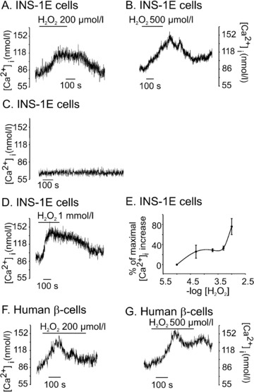
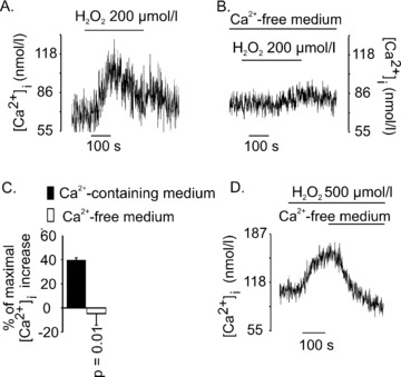
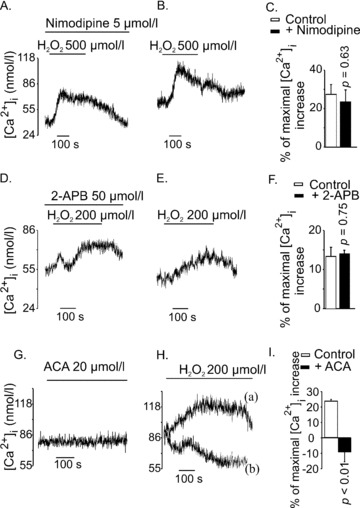
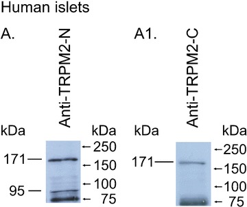
Type 2 melastatin-related transient receptor potential channel (TRPM2), a member of the melastatin-related TRP (transient receptor potential) subfamily is a Ca(2+)-permeable channel activated by hydrogen peroxide (H(2)O(2)). We have investigated the role of TRPM2 channels in mediating the H(2)O(2)-induced increase in the cytoplasmic free Ca(2+) concentration ([Ca(2+)](i)) in insulin-secreting cells. In fura-2 loaded INS-1E cells, a widely used model of beta-cells, and in human beta-cells, H(2)O(2) increased [Ca(2+)](i), in the presence of 3 mM glucose, by inducing Ca(2+) influx across the plasma membrane. H(2)O(2)-induced Ca(2+) influx was not blocked by nimodipine, a blocker of the L-type voltage-gated Ca(2+) channels nor by 2-aminoethoxydiphenyl borate, a blocker of several TRP channels and store-operated channels, but it was completely blocked by N-(p-amylcinnamoyl)anthranilic acid (ACA), a potent inhibitor of TRPM2. Adenosine diphosphate phosphate ribose, a specific activator of TRPM2 channel and H(2)O(2), induced inward cation currents that were blocked by ACA. Western blot using antibodies directed to the epitopes on the N-terminal and on the C-terminal parts of TRPM2 identified the full length TRPM2 (TRPM2-L), and the C-terminally truncated TRPM2 (TRPM2-S) in human islets. We conclude that functional TRPM2 channels mediate H(2)O(2)-induced Ca(2+) entry in beta-cells, a process potently inhibited by ACA.


Figures

Figure 1
Figure 1
H2O2 increased [Ca2+]i in INS-1E cells and human β-cells. Cells were perfused with physiological solution containing 3 mM glucose and [Ca2+]i was measured in fura-2 loaded cells by microfluorometry. (A) H2O2 (200 μM) increased [Ca2+]i in a reversible manner (n= 16). (B) and (D) [Ca2+]i increase by 500 μM, (n= 24) and 1 mM H2O2, (n= 9) did not completely reverse after washout of H2O2. (C) baseline [Ca2+]i remained stable when no H2O2 was added. (E) the concentration-response curve for H2O2-induced [Ca2+]i increase. Data are expressed as percentage of maximal [Ca2+]i increase obtained by 1 mM H2O2. Each point represents a mean of 5–24 experiments. (F) and (G) show the effects of H2O2 on [Ca2+]i in single human β-cells. H2O2 increased [Ca2+]i in 10 out of 12 cells for 200 μM and 4 out of 6 for 500 μM.
Figure 2
Figure 2
H2O2-induced [Ca2+]i increase was due to Ca2+ entry across the plasma membrane. H2O2 (200 μM) was applied to INS-1E cells in the presence of 1.5 mM extracellular Ca2+ (A) or in nominally Ca2+-free medium (B). Maximal [Ca2+]i change in Ca2+ containing medium was 39±2 nM (n= 3) and that in nominally Ca2+-free medium was –5±10 nM (n= 3) (C). In (D), cells were first exposed to H2O2 (500 μM) in the presence of 1.5 mM extracellular Ca2+, and the solution was switched to a nominally Ca2+-free medium at the time indicated by the horizontal bar(n= 5) (c.f.Fig. 1B).
Figure 3
Figure 3
Effects of different channel blockers on H2O2-induced [Ca2+]i increase in INS-1E cells. (A) and (B) Nimodipine did not inhibit H2O2-induced [Ca2+]i response. Cells were pre-incubated with Nimodipine (5 μM) for 10 min. H2O2 (500 μM) was applied in the continued presence of nimodipine (the trace is representative of seven independent experiments). (B) Shows control experiments for (A) (the trace is representative of eight independent experiments). (D) and (E) 2-APB (50 μM) did not alter the [Ca2+]i response to H2O2 (200 μM). Cells were treated with 2-APB (50 μM) for 10 min. during pre-incubation and H2O2 was applied in the continued presence of 2-APB. (E) The control for (A) showing [Ca2+]i response to H2O2 in the absence of 2-APB (traces D and E are representatives of three to five independent experiments). (G) ACA (20 μM) did not increase [Ca2+]i in INS-1E cells. (H) ACA (20 μM) completely inhibited H2O2-induced [Ca2+]i response. Trace (a) shows [Ca2+]i response to H2O2 (200 μM) in the absence of ACA (the trace is representative of three independent experiments). In trace (b), ACA (20 μM) was applied 1 min. before application of H2O2 (200 μM) and was continuously present in the perfusion (the trace is representative of three independent experiments). (I) Maximal Ca2+ changes by H2O2 in controls and in the ACA-treated cells were 23 ± 1 and –9 ± 6 nM, respectively (P < 0.01, n= 6). In (C), (F) and (I), the bars represent mean [Ca2+]i increase obtained by H2O2, expressed as the percentage of maximal [Ca2+]i increase obtained by 25 mM KCl in respective experiments.
Figure 4
Figure 4
Whole-cell currents induced by ADP ribose and H2O2 in INS-1E cells. The whole-cell configuration was attained at the point indicated with ‘w.c.’. Recordings were performed at RT and the holding potential was –60 mV. Bars indicate times where the standard bath solution was changed to a solution containing either NMDG+ or the TRPM2 channel inhibitor ACA. (A) Whole-cell current recorded in the presence of intracellular ADP ribose. The pipette solution contained 0.6 mM ADP ribose and 1 μM Ca2+. (B) Whole cell currents recorded without ADP ribose and after application of 1–2 μl 30% H2O2 directly into the recording chamber. The estimated final concentration of H2O2 in the chamber was ∼10 mM. The pipette solution contained 1 μM free Ca2+. (C) Current–voltage relationship of H2O2-induced currents as derived from (B), recorded during voltage ramps from –90 to +60 mV of 400 ms duration.
Figure 5
Figure 5
Western blot analysis of TRPM2 proteins in human islets. A total of 90 μg of membrane proteins from human islets were separated in two lanes (A, A1) by 10% SDS-PAGE electrophoresis. In A, the blots were probed with an anti-TRPM2-N antibody (1:300). In A1, the blots were probed with an anti-TRPM2-C antibody (1:300). Standard proteins are shown on the right side. The experiments have been repeated at least three times with similar results.

References

    1. Perraud AL, Fleig A, Dunn CA, et al. ADP-ribose gating of the calcium-permeable LTRPC2 channel revealed by Nudix motif homology. Nature. 2001;411:595–9. - PubMed
    1. Maechler P, Jornot L, Wollheim CB. Hydrogen peroxide alters mitochondrial activation and insulin secretion in pancreatic beta cells. J Biol Chem. 1999;274:27905–13. - PubMed
    1. Pi J, Bai Y, Zhang Q, et al. Reactive oxygen species as a signal in glucose-stimulated insulin secretion. Diabetes. 2007;56:1783–91. - PubMed
    1. Qian F, Huang P, Ma L, et al. TRP genes: candidates for nonselective cation channels and store-operated channels in insulin-secreting cells. Diabetes. 2002;51:S183–9. - PubMed
    1. Togashi K, Hara Y, Tominaga T, et al. TRPM2 activation by cyclic ADP-ribose at body temperature is involved in insulin secretion. EMBO J. 2006;25:1804–15. - PMC - PubMed

Publication types