

Complex patterns of mitochondrial dynamics in human pancreatic cells revealed by fluorescent confocal imaging
- PMID: 19382913
- PMCID: PMC3837585
- DOI: 10.1111/j.1582-4934.2009.00750.x
Complex patterns of mitochondrial dynamics in human pancreatic cells revealed by fluorescent confocal imaging
Abstract
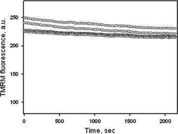
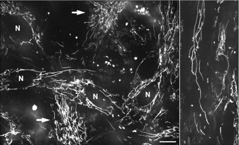
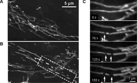
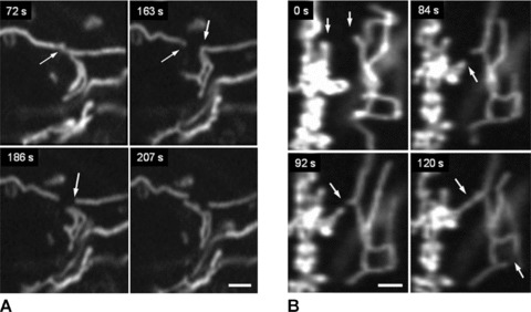
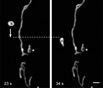
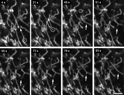
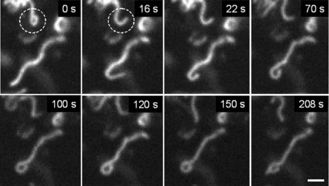
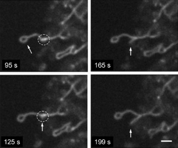
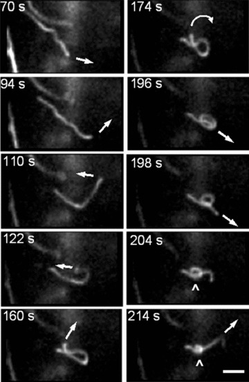
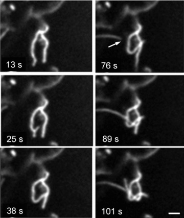
Mitochondrial morphology and intracellular organization are tightly controlled by the processes of mitochondrial fission-fusion. Moreover, mitochondrial movement and redistribution provide a local ATP supply at cellular sites of particular demands. Here we analysed mitochondrial dynamics in isolated primary human pancreatic cells. Using real time confocal microscopy and mitochondria-specific fluorescent probes tetramethylrhodamine methyl ester and MitoTracker Green we documented complex and novel patterns of spatial and temporal organization of mitochondria, mitochondrial morphology and motility. The most commonly observed types of mitochondrial dynamics were (i) fast fission and fusion; (ii) small oscillating movements of the mitochondrial network; (iii) larger movements, including filament extension, retraction, fast (0.1-0.3 mum/sec.) and frequent oscillating (back and forth) branching in the mitochondrial network; (iv) as well as combinations of these actions and (v) long-distance intracellular translocation of single spherical mitochondria or separated mitochondrial filaments with velocity up to 0.5 mum/sec. Moreover, we show here for the first time, a formation of unusual mitochondrial shapes like rings, loops, and astonishingly even knots created from one or more mitochondrial filaments. These data demonstrate the presence of extensive heterogeneity in mitochondrial morphology and dynamics in living cells under primary culture conditions. In summary, this study reports new patterns of morphological changes and dynamic motion of mitochondria in human pancreatic cells, suggesting an important role of integrations of mitochondria with other intracellular structures and systems.
Figures
Similar articles
-
The cell-type specificity of mitochondrial dynamics.Int J Biochem Cell Biol. 2009 Oct;41(10):1928-39. doi: 10.1016/j.biocel.2009.03.007. Epub 2009 Mar 27. Int J Biochem Cell Biol. 2009. PMID: 19703655 Review.
-
Altered mitochondrial structure and motion dynamics in living cells with energy metabolism defects revealed by real time microscope imaging.Microsc Microanal. 2004 Apr;10(2):247-60. doi: 10.1017/S143192760404005X. Microsc Microanal. 2004. PMID: 15306050
-
A faster, high resolution, mtPA-GFP-based mitochondrial fusion assay acquiring kinetic data of multiple cells in parallel using confocal microscopy.J Vis Exp. 2012 Jul 20;(65):e3991. doi: 10.3791/3991. J Vis Exp. 2012. PMID: 22847388 Free PMC article.
-
Understanding Mitochondrial and Lysosomal Dynamics by Fluorescent Microscopy.Methods Mol Biol. 2025;2878:211-221. doi: 10.1007/978-1-0716-4264-1_11. Methods Mol Biol. 2025. PMID: 39546264
-
Dynamics of mitochondria in living cells: shape changes, dislocations, fusion, and fission of mitochondria.Microsc Res Tech. 1994 Feb 15;27(3):198-219. doi: 10.1002/jemt.1070270303. Microsc Res Tech. 1994. PMID: 8204911 Review.
Cited by
-
An improved quantitative approach for the assessment of mitochondrial fragmentation in chemoresistant ovarian cancer cells.PLoS One. 2013 Sep 9;8(9):e74008. doi: 10.1371/journal.pone.0074008. eCollection 2013. PLoS One. 2013. PMID: 24040144 Free PMC article.
-
Mitochondrial network expansion and dynamic redistribution during islet morphogenesis in zebrafish larvae.FEBS Lett. 2023 Jan;597(2):262-275. doi: 10.1002/1873-3468.14508. Epub 2022 Oct 19. FEBS Lett. 2023. PMID: 36217213 Free PMC article.
-
Analysis of Mitochondrial Function, Structure, and Intracellular Organization In Situ in Cardiomyocytes and Skeletal Muscles.Int J Mol Sci. 2022 Feb 18;23(4):2252. doi: 10.3390/ijms23042252. Int J Mol Sci. 2022. PMID: 35216368 Free PMC article. Review.
-
Real-Time Live Confocal Fluorescence Microscopy as a New Tool for Assessing Platelet Vitality.Transfus Med Hemother. 2010;37(5):299-305. doi: 10.1159/000320368. Epub 2010 Sep 15. Transfus Med Hemother. 2010. PMID: 21113254 Free PMC article.
-
Single cell optical imaging and spectroscopy.Chem Rev. 2013 Apr 10;113(4):2469-527. doi: 10.1021/cr300336e. Epub 2013 Feb 14. Chem Rev. 2013. PMID: 23410134 Free PMC article. Review. No abstract available.
References
-
- McBride HM, Neuspiel M, Wasiak S. Mitochondria: more than just a powerhouse. Curr Biol. 2006;16:R551–60. - PubMed
-
- Newmeyer DD, Ferguson-Miller S. Mitochondria: Releasing power for life and unleashing the machineries of death. Cell. 2003;112:873. - PubMed
-
- Kroemer G, Reed JC. Mitochondrial control of cell death. Nat Med. 2000;6:513–9. - PubMed
Publication types
MeSH terms
Substances
LinkOut - more resources
Full Text Sources
