

Expression levels of immune markers in Actinobacillus pleuropneumoniae infected pigs and their relation to breed and clinical symptoms
- PMID: 19383119
- PMCID: PMC2678107
- DOI: 10.1186/1746-6148-5-13
Expression levels of immune markers in Actinobacillus pleuropneumoniae infected pigs and their relation to breed and clinical symptoms
Abstract
Background: In pigs little is known about the role of innate immune defence in bacterial infections of the respiratory tract, despite their major role in pig production. In the present study we characterized and compared in vitro and in vivo activation of immune markers of different pig breeds 7 days before, and 4 and 21 days after an experimental aerosol infection with Actinobacillus (A.) pleuropneumoniae.
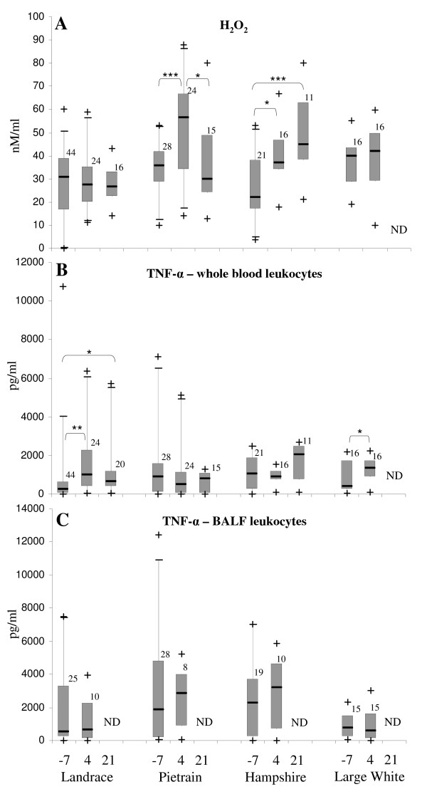
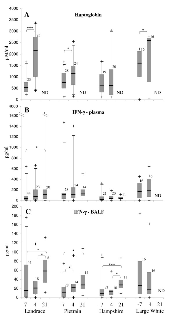
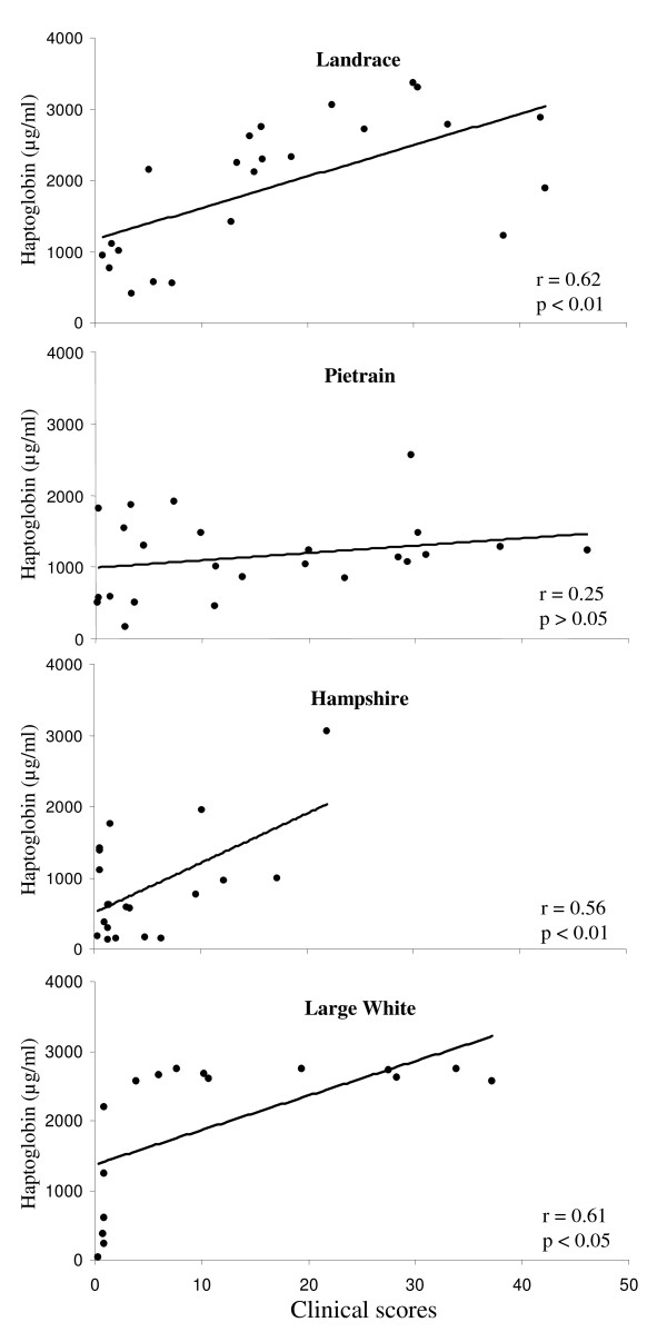
Results: In vitro stimulation of bronchoalveolar lavage fluid (BALF) and blood leukocytes with A. pleuropneumoniae, Streptococcus suis, PMA and LPS led to production of different amounts of H2O2, NO and TNF-alpha, depending on the stimulus, individual, breed and time of infection. Generally, significant responses to in vitro stimulation were observed only in blood leukocytes, whereas the alveolar macrophages showed a high basal activation. In addition, the production of haptoglobin and cytokines (TNF-alpha, IFN-gamma and IL-10) in vivo was measured in plasma and BALF. Plasma haptoglobin levels mirrored the clinical manifestations at 4 days post-infection. In plasma and BALF TNF-alpha could not be detected, whereas variable levels of IFN-gamma were found at pre- and post-infection times. IL-10 was found in some plasma but in none of the BALF samples. The different expression levels in individuals within the breeds correlated for some markers with the severity of clinical manifestations, e.g. H2O2, plasma haptoglobin and BALF IFN-gamma for German Landrace pigs.
Conclusion: Our findings revealed differences in the activation of the immune markers with respect to infection time, individuals and breeds. Moreover, results showed different correlation grades between the immune markers produced in vitro or in vivo and the clinical manifestations. Further analyses will have to show whether these markers may serve as correlates of protection against porcine respiratory infections.
Figures
References
-
- Sebunya TNK, Saunders JR. Haemophilus pleuropneumoniae infection in swine: a review. J Am Vet Med Assoc. 1983;182:1331–1337. - PubMed
-
- Gobert AP, Wilson KT, Martin C. Cellular responses to attaching and effacing bacteria: activation and implication of the innate immune system. Arch Immunol Ther Exp (Warsz) 2005;53:234–244. - PubMed
-
- Hodgson JC, Watkins CA, Bayne CW. Contribution of respiratory burst activity to innate immune function and the effects of disease status and agent on chemiluminescence responses by ruminant phagocytes in vitro. Vet Immunol Immunopathol. 2006;112:12–23. doi: 10.1016/j.vetimm.2006.03.008. - DOI - PubMed
Publication types
MeSH terms
Substances
LinkOut - more resources
Full Text Sources
