

Imaging and cancer: a review
- PMID: 19383333
- PMCID: PMC5527766
- DOI: 10.1016/j.molonc.2008.04.001
Imaging and cancer: a review
Erratum in
-
Corrigendum to: Imaging and cancer: a review.Mol Oncol. 2022 Aug;16(15):2896. doi: 10.1002/1878-0261.13283. Mol Oncol. 2022. PMID: 35921212 Free PMC article. No abstract available.
Abstract
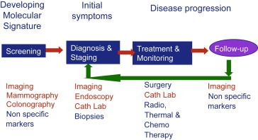
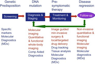
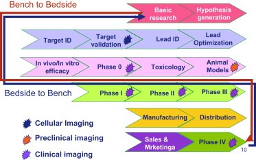
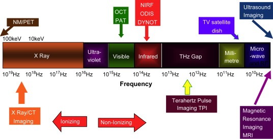
Multiple biomedical imaging techniques are used in all phases of cancer management. Imaging forms an essential part of cancer clinical protocols and is able to furnish morphological, structural, metabolic and functional information. Integration with other diagnostic tools such as in vitro tissue and fluids analysis assists in clinical decision-making. Hybrid imaging techniques are able to supply complementary information for improved staging and therapy planning. Image guided and targeted minimally invasive therapy has the promise to improve outcome and reduce collateral effects. Early detection of cancer through screening based on imaging is probably the major contributor to a reduction in mortality for certain cancers. Targeted imaging of receptors, gene therapy expression and cancer stem cells are research activities that will translate into clinical use in the next decade. Technological developments will increase imaging speed to match that of physiological processes. Targeted imaging and therapeutic agents will be developed in tandem through close collaboration between academia and biotechnology, information technology and pharmaceutical industries.
Figures
References
-
- Aberg, P. , Nicander, I. , Hansson, J. , Geladi, P. , Holmgren, U. , Ollmar, S. , 2004. Skin cancer identification using multifrequency electrical impedance – a potential screening tool. IEEE Transactions on Biomedical Engineering 51, 2097–2102. - PubMed
-
- Aboagye, E.O. , Bhujwalla, Z.M. , 1999. Malignant transformation alters membrane choline phospholipid metabolism of human mammary epithelial cells. Cancer Research 59, 80–84. - PubMed
-
- Aboagye, E.O. , Bhujwalla, Z.M. , Shungu, D.C. , Glickson, J.D. , 1998. Detection of tumour response to chemotherapy by 1H nuclear magnetic resonance spectroscopy: effect of 5-fluorouracil on lactate levels in radiation-induced fibrosarcoma I tumours. Cancer Research 58, 1063–1067. - PubMed
-
- Abourbeh, G. , Dissoki, S. , Jacobson, O. , 2007. Evaluation of radiolabeled ML04, a putative irreversible inhibitor of epidermal growth factor receptor, as a bioprobe for PET imaging of EGFR-overexpressing tumors. Nuclear Medicine and Biology 34, 55–70. - PubMed
Publication types
MeSH terms
LinkOut - more resources
Full Text Sources
Other Literature Sources
Medical
