. 2008 Oct;2(3):223-32.
doi: 10.1016/j.molonc.2008.06.002. Epub 2008 Jun 18.

Estradiol-estrogen receptor: a key interplay of the expression of syndecan-2 and metalloproteinase-9 in breast cancer cells

Affiliations

Estradiol-estrogen receptor: a key interplay of the expression of syndecan-2 and metalloproteinase-9 in breast cancer cells

Olga Ch Kousidou et al. Mol Oncol. 2008 Oct.

Abstract

Estrogens are related with the growth and development of target tissues and play a critical role in breast cancer progression. The effects of estrogens are mediated by the estrogen receptors ERalpha and ERbeta, which are members of the nuclear steroid receptor superfamily. To date, it is not known how these hormones elicit many of their effects on extracellular matrix molecules and how these effects can be connected with ER expression. For this purpose, the effect of estradiol on ER expression as well as on proteoglycan and metalloproteinase expression was studied. The effect of E2 on extracellular matrix molecule expression has been studied using ERalpha suppression in breast cancer cells. Our studies using ERalpha-positive MCF-7 cells show that estradiol affects the expression of syndecan-2, but not of syndecan-4, through ERalpha. Furthermore, the ability of estradiol to affect MMP-9 and TIMP-1 expression is connected with ERalpha status. Together, these data demonstrate the significant role of ERalpha on mediating the effect of estradiol on extracellular matrix molecules.


Figures

Figure 1
Figure 1
Estradiol effects on the expression of ERα and ERβ in three cell lines (MCF‐7, MDA‐MB‐231 and ZR‐75‐1). (A), (B) show the effect of estradiol on gene expression of ERs, while (C), (D) show its effect on protein level. Statistically significant differences among the estradiol‐treated and control cells are shown by asterisks (*p≤0.01). RT–PCR analyses are shown in insets.
Figure 2
Figure 2
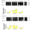
Effect of estradiol on the expression of syndecans‐2 and ‐4, in three cell lines (MCF‐7, MDA‐MB‐231 and ZR‐75‐1). The results are expressed as the average±SD of three experiments in triplicate. Statistically significant differences among the estradiol‐treated and control cells are shown by asterisks (*p≤0.01). RT–PCR analyses are shown in insets.
Figure 3
Figure 3
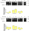
Effect of estradiol on the expression of MMP‐9 and TIMP‐1, in three cell lines (MCF‐7, MDA‐MB‐231 and ZR‐75‐1). The results are expressed as the average±SD of three experiments in triplicate. Statistically significant differences among the estradiol‐treated and control cells are shown by asterisks (*p≤0.01). RT–PCR analyses are shown in insets.
Figure 4
Figure 4
Time‐dependent suppression of ERα in MCF‐7 cells. Cells were transfected using the Lipofectamine reagent according to manufacturers’ instructions. The levels of ERα in control, scramble and siRNA treated cells were quantified by real‐time PCR. Results are expressed as the mean±SD of three independent experiments. Statistically significant differences among MCF‐7 scramble and after transfection cells are shown by asterisks.
Figure 5
Figure 5
Effect of estradiol on the expression of syndecan‐2 and ‐4 of ERα‐suppressed MCF‐7 cells. MCF‐7 cells were transfected with scrambled (A, C) or ERα (B, D) siRNA and the levels of syndecan‐2 and ‐4 were quantified by real‐time PCR. Results are expressed as the mean±SD of three independent experiments. Statistically significant differences among MCF‐7 scramble and after transfection cells are shown by asterisks.
Figure 6
Figure 6
Effect of estradiol on the expression of MMP‐9 and TIMP‐1 of ERα‐suppressed MCF‐7 cells. MCF‐7 cells were transfected with scrambled (A, C) or ERα (B, D) siRNA and the levels of MMP‐9 and TIMP‐1 were quantified by real‐time PCR. Results are expressed as the mean±SD of three independent experiments. Statistically significant differences among MCF‐7 scramble and after transfection cells are shown by asterisks.

References

    1. Alarid, E.T. , Bakopoulos, N. , Solodin, N. , 1999. Proteasome-mediated proteolysis of estrogen receptor: a novel component in autologous down-regulation. Mol. Endocrinol 13, 1522–1534. - PubMed
    1. Ali, S. , Coombes, R.C. , 2000. Estrogen receptor alpha in human breast cancer: occurrence and significance. J. Mammary Gland Biol. Neoplasia 5, 271–281. - PubMed
    1. Beauvais, D.L.M. , Rapraeger, A. , 2004. Syndecans in tumor cell adhesion and signaling. Reprod. Biol. Endocrinol 2, 1–12. - PMC - PubMed
    1. Borras, M. , Hardy, L. , Lempereur, F. , El Khissiin, A.H. , Legros, N. , Gol-Winkler, R. , Leclercq, G. , 1994. Estradiol-induced down-regulation of estrogen receptor. Effect of various modulators of protein synthesis and expression. J. Steroid Biochem. Mol. Biol. 48, 325–336. - PubMed
    1. Capony, F. , Morisset, M. , Barrett, A.J. , Capony, J.P. , Broquet, P. , Vignon, F. , Chambon, M. , Louisot, P. , Rochefort, H. , 1987. Phosphorylation, glycosylation, and proteolytic activity of the 52-kD estrogen-induced protein secreted by MCF-7 cells. J. Cell Biol. 104, 253–262. - PMC - PubMed