. 2009 Apr 22;96(8):3116-25.
doi: 10.1016/j.bpj.2009.01.021.

Transitions between closed and open conformations of TolC: the effects of ions in simulations

Affiliations

Transitions between closed and open conformations of TolC: the effects of ions in simulations

Robert Schulz et al. Biophys J. .

Abstract

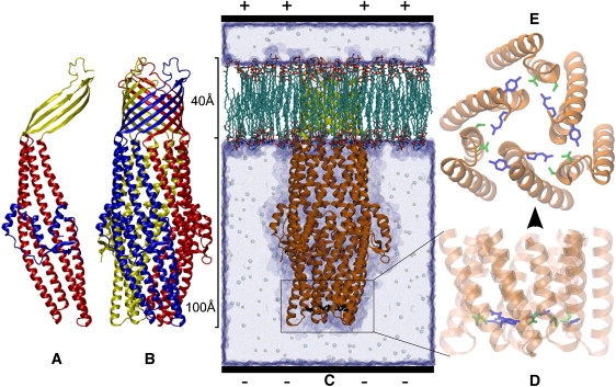
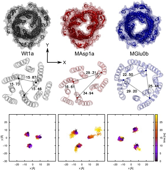
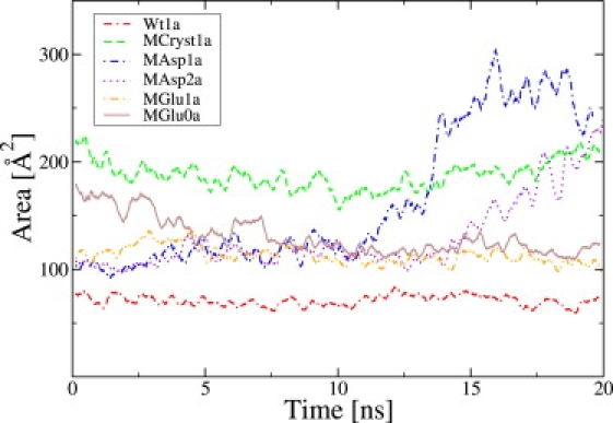
Bacteria, such as Escherichia coli, use multidrug efflux pumps to export toxic substrates through their cell membranes. Upon formation of an efflux pump, the aperture of its outer membrane protein TolC opens and thereby enables the extrusion of substrate molecules. The specialty of TolC is its ability to dock to different transporters, making it a highly versatile export protein. Within this study, the transition between two conformations of TolC that are both available as crystal structures was investigated using all-atom molecular dynamics simulations. To create a partially open conformation from a closed one, the stability of the periplasmic aperture was weakened by a double point mutation at the constricting ring, which removes some salt bridges and hydrogen bonds. These mutants, which showed partial opening in previous experiments, did not spontaneously open during a 20-ns equilibration at physiological values of the KCl solution. Detailed analysis of the constricting ring revealed that the cations of the solvent were able to constitute ionic bonds in place of the removed salt bridges, which inhibited the opening of the aperture in simulations. To remove the ions from these binding positions within the available simulation time, an extra force was applied onto the ions. To keep the effect of this additional force rather flexible, it was applied in form of an artificial external electric field perpendicular to the membrane. Depending on the field direction and the ion concentration, these simulations led to a partial opening. In experiments, this energy barrier for the ions can be overcome by thermal fluctuations on a longer timescale.


Figures

Figure 1
Figure 1
TolC protein structure in cartoon representation of the secondary structure (50) with (A) the β-barrel above and α-helical coiled coils below. (B) The monomers in different colors. (C) The protein in a simulation box with the orientation of the positive applied voltage (see Simulation Setup). (D) Zoom to the aperture with the constricting ring in licorice representation of the side chains (dark shaded, T152 and D153; light shaded, Y362 and R367). (E) Side view of panel D looking from the periplasm toward the membrane part (figures created with VMD (40)).
Figure 2
Figure 2
Results of simulations with applied voltage: In the first row, snapshots of the protein with backbone (cartoon) and all-atom (transparent surface) representation of the last frame are drawn using the same perspective as in Fig. 1E. In the second line, the distances (in Å) between the Cα atoms (van der Waals representation) of G365 are displayed with the protein backbone in transparent cartoon representation. In the third line, the traces of the residue G365 Cα atoms are shown with time-dependent coloring, starting from the equilibration.
Figure 3
Figure 3
Porcupine plot pointing from open crystal structure (2VDE) to MGlu0b final structure with arrows per residue Cα atom lengthened and colored due to deviation value (values <2 Å omitted). (Top) Direction of view: perpendicular to the membrane; (bottom): toward the membrane (see Fig. 1).
Figure 4
Figure 4
Comparison of the triangular area. The shown results are for an external voltage of +1 V. A running average has been calculated for each 250 ps.
Figure 5
Figure 5
The same as in Fig. 4, but for a voltage of −1 V.
Figure 6
Figure 6
Slices through the potential map for different simulations: (A) Wt1a, (B) MGlu1a, (C) MAsp1a, and (D) MAsp1b.
Figure 7
Figure 7
The MGlu aperture (cartoon representation) with the residue side chains (licorice representation colored by amino-acid type) of the cation pockets (transparent surface representation) occupied with potassium ions (van der Waals representation). Same perspective as in Fig. 1E. Each cation pocket consists of the residues T152, D153, and E367.
Figure 8
Figure 8
Occupation for each of the three cation pockets with ion selection range of 3 Å. A black line represents that the respective pocket is occupied.
Figure 9
Figure 9
Average occupation frequency of the cation pockets. The label “equi” denotes the field-free equilibration runs, and the letters a, b, and c stand for +1 V, −1 V, and +0.5 V, respectively.

References

    1. Fernandes P. Antibacterial discovery and development—the failure of success? Nat. Biotechnol. 2006;24:1497–1503. - PubMed
    1. Piddock L. Clinically relevant chromosomally encoded multidrug resistance efflux pumps in bacteria. Clin. Microbiol. Rev. 2006;19:382–402. - PMC - PubMed
    1. Lomovskaya O., Zgurskaya H.I., Totrov M., Watkins W.J. Waltzing transporters and “the dance macabre” between humans and bacteria. Nat. Rev. Drug Discov. 2007;6:56–65. - PubMed
    1. Alberts B. 2nd Ed. Garland Science; New York: 2004. Essential Cell Biology.
    1. Alberts B., Johnson A., Lewis J., Raff M., Roberts K. 4th Ed. Garland Science; New York: 2002. Molecular Biology of the Cell.

MeSH terms

Substances

LinkOut - more resources