. 2009 Apr 22;96(8):3197-209.
doi: 10.1016/j.bpj.2009.01.026.

Structure of self-aggregated alamethicin in ePC membranes detected by pulsed electron-electron double resonance and electron spin echo envelope modulation spectroscopies

Affiliations

Structure of self-aggregated alamethicin in ePC membranes detected by pulsed electron-electron double resonance and electron spin echo envelope modulation spectroscopies

Alexander D Milov et al. Biophys J. .

Abstract

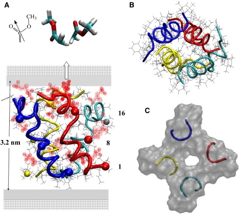
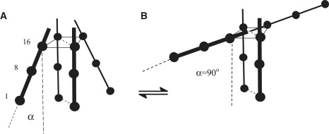
PELDOR spectroscopy was exploited to study the self-assembled super-structure of the [Glu(OMe)(7,18,19)]alamethicin molecules in vesicular membranes at peptide to lipid molar ratios in the range of 1:70-1:200. The peptide molecules were site-specifically labeled with TOAC electron spins. From the magnetic dipole-dipole interaction between the nitroxides of the monolabeled constituents and the PELDOR decay patterns measured at 77 K, intermolecular-distance distribution functions were obtained and the number of aggregated molecules (n approximately 4) was estimated. The distance distribution functions exhibit a similar maximum at 2.3 nm. In contrast to Alm16, for Alm1 and Alm8 additional maxima were recorded at 3.2 and approximately 5.2 nm. From ESEEM experiments and based on the membrane polarity profiles, the penetration depths of the different spin-labeled positions into the membrane were qualitatively estimated. It was found that the water accessibility of the spin-labels follows the order TOAC-1 > TOAC-8 approximately TOAC-16. The geometric data obtained are discussed in terms of a penknife molecular model. At least two peptide chains are aligned parallel and eight ester groups of the polar Glu(OMe)(18,19) residues are suggested to stabilize the self-aggregate superstructure.


Figures

Figure 1
Figure 1
Continuous wave ESR spectra of spin-labeled alamethicin analogs at 77 K. Curve 1: Alm1 (2 × 10⁻³ M) in methanol (containing 5% ethanol). Curves 2–4: Alm1, Alm8, and Alm16 in ePC vesicles at a P/L molar ratio of 1:150.
Figure 2
Figure 2
(A) PELDOR signal decays for spin-labeled alamethicin analogs bound to the membranes of ePC vesicles at 77 K. The P/L molar ratio is 1:70 for curves 1, 3, and 4, and 1:160 for curve 2. Curves 1 and 2 are shifted downward by 0.4 and curve 3 is shifted downward by 0.2. (B) VINTRA signal decays for frozen solutions of alamethicin analogs in ePC vesicles.
Figure 3
Figure 3
Distance distribution functions F(r) between spin-labels for alamethicin analogs in membranes of frozen ePC vesicles. Curves 2 and 3 are shifted upward by 0.5 and 1.0, respectively. The distance distributions are obtained from the experimental VINTRA decays by the Tikhonov regularization method. The shape of F(r) at larger distances than that indicated by the arrow is not authentic and can be used for the estimation of the area in this r-region.
Figure 4
Figure 4
(A) ESEEM decays for Alm1 bound to membranes of D2O-hydrated ePC vesicles. The stimulated echo signal (V) dependence оn the delay between the first and the third pulses, t, is shown in curve 1. Curve 2 is smoothed by a six-order polynomial. (B) The normalized spin echo dependence on the delay time t for spin-labeled alamethicin analogs in ePC vesicles hydrated in D2O buffer. The P/L molar ratio is 1:70. The spin echo signal Vn is normalized.
Figure 5
Figure 5
Energy minimized model of the supramolecular alamethicin tetramer, which is based on the short 2.3 nm PELDOR distance between the exocyclic oxygen atoms of the TOAC-1, TOAC-8, and TOAC-16 spin-labels. The spatial positions of the oxygen radicals are indicated by balls, with the TOAC-1 labels shown at the bottom of the figure. For clarity, but in contrast to our experiments, each peptide is threefold labeled. (A) A side view of the tube model shows the spatial arrangement of four parallel aligned α-helical peptide chains, which make angles of −15° (blue and red chains), +13° (cyan chain), and −8° (yellow chain), with respect to the membrane normal. The helices are bent at the Leu12-Aib13 sequence due to the missing hydrogen bonds between the carbonyl oxygens of the Gly11 and the tertiary nitrogen atoms of the Pro14 residues. The maximum diameter of the peptide complex (3.2 nm) appears roughly matching the hydrophobic thickness of the membrane double layer (3.5 nm). For convenience, the zwitterionic and polar regions of the membrane are also depicted. The locations of the flexible polar side chains of the γ-ester groups of the Glu(OMe)7,18,19 residues are shown by dotted balls. A more detailed arrangement, particularly for the (Glu(OMe)18 residues of the red and cyan colored peptide chains, is shown in the inset of A. These mutually antiparallel oriented γ-ester groups are located at a distance of 0.36 nm. The electric dipole-dipole interactions between the ester groups are believed to stabilize the C-terminal ends of the peptide chains. This stable region of the peptide complex might be further supported by the relative narrow PELDOR distribution of distances found for TOAC-16. (B and C) Top down views of the peptide complex (the N-terminal ends are shown at the front side). (B) Tube model of the spatial arrangement of the helical chains. (C) A semitransparent surface model of a thin slice with a thickness of about half of a helix-turn and showing the entry of one or more channels within the tetramer.
Figure 6
Figure 6
(A) Schematic representation of the model characterized by a penknife type of association, i.e., two parallel aligned transmembrane peptides, whereas two other peptide chains are making an angle α. Distances between the TOAC-16 labels, indicated by solid lines, are kept fixed at 2.3 nm. The positions of the spin-labels along the schematic peptide chains are shown by balls. (B) The T-shaped tetramer structure, defined by α = 90°, qualitatively explains the occurrence of both the short and long experimental distances between the TOAC-1 and TOAC-8 labels of 2.3 + 3.2 nm and 5 nm, whereas for TOAC-16 a distance of ∼2.3 nm only was found. Together with the water accessibility of the labels estimated by ESEEM spectroscopy, the combined data might be understood by considering the presence of two types of aggregated species, corresponding to α = 0° (transmembrane tetramer) and α = 90° (T-shaped complex).
None

Similar articles

Cited by

References

    1. Leitgeb B., Szekeres L., Manczinger L., Vágvölgyi C., Kredics L. The history of alamethicin: a review of the most extensively studied peptaibol. Chem. Biodivers. 2007;4:1027–1051. - PubMed
    1. Mueller P., Rudin P.O. Action potentials induced in biomolecular lipid membranes. Nature. 1968;217:713–719. - PubMed
    1. Boheim G. Statistical analysis of alamethicin in a lipid membrane. J. Membr. Biol. 1974;19:277–303. - PubMed
    1. Sansom M.S.P. Alamethicin and related peptaibols—model ion channels. Eur. Biophys. J. 1993;22:105–124. - PubMed
    1. Baumann G., Mueller P. A molecular model of membrane excitability. J. Supramol. Struct. 1974;2:538–577. - PubMed

Publication types

LinkOut - more resources