. 2009 Apr 22;96(8):3305-18.
doi: 10.1016/j.bpj.2008.12.3960.

Surface-bound casein modulates the adsorption and activity of kinesin on SiO2 surfaces

Affiliations

Surface-bound casein modulates the adsorption and activity of kinesin on SiO2 surfaces

Tomomitsu Ozeki et al. Biophys J. .

Abstract

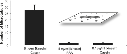
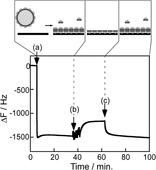
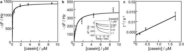
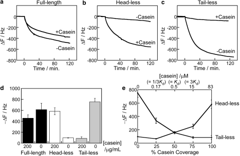
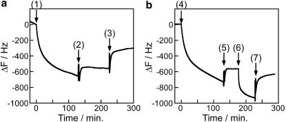
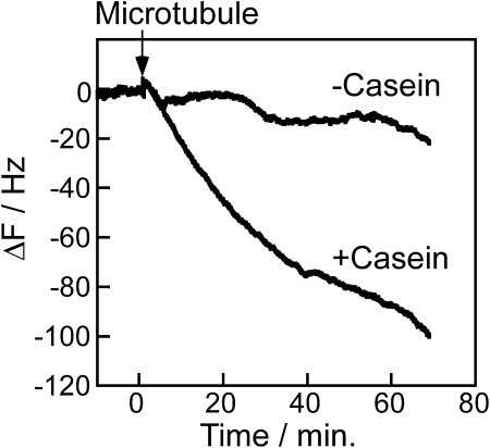
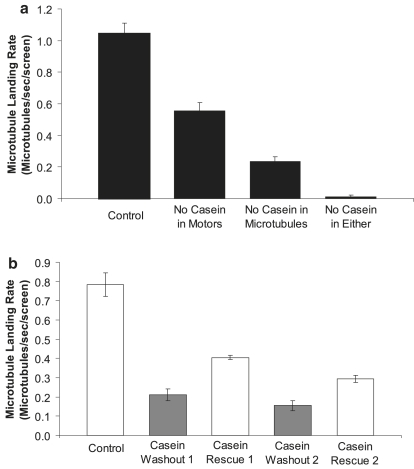
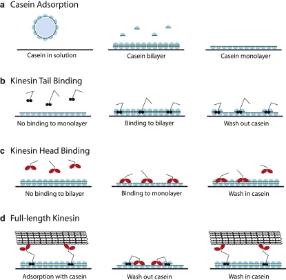
Conventional kinesin is routinely adsorbed to hydrophilic surfaces such as SiO(2). Pretreatment of surfaces with casein has become the standard protocol for achieving optimal kinesin activity, but the mechanism by which casein enhances kinesin surface adsorption and function is poorly understood. We used quartz crystal microbalance measurements and microtubule gliding assays to uncover the role that casein plays in enhancing the activity of surface-adsorbed kinesin. On SiO(2) surfaces, casein adsorbs as both a tightly bound monolayer and a reversibly bound second layer that has a dissociation constant of 500 nM and can be desorbed by washing with casein-free buffer. Experiments using truncated kinesins demonstrate that in the presence of soluble casein, kinesin tails bind well to the surface, whereas kinesin head binding is blocked. Removing soluble casein reverses these binding profiles. Surprisingly, reversibly bound casein plays only a moderate role during kinesin adsorption, but it significantly enhances kinesin activity when surface-adsorbed motors are interacting with microtubules. These results point to a model in which a dynamic casein bilayer prevents reversible association of the heads with the surface and enhances association of the kinesin tail with the surface. Understanding protein-surface interactions in this model system should provide a framework for engineering surfaces for functional adsorption of other motor proteins and surface-active enzymes.


Figures

Figure 1
Figure 1
Comparing the microtubule-binding activity of kinesin in casein and BSA. In the casein experiment, flow cells were pretreated with 0.5 mg/mL casein, and 0.2 mg/mL casein was present in both the kinesin and microtubule solutions. In the BSA experiment, casein was replaced by 0.5 mg/mL BSA in all solutions. Five minutes after introduction of rhodamine-labeled microtubules (64 nM), the number of microtubules moving in each 70 μm × 55 μm video screen was counted. Data for each condition are plotted as mean ± SD for N = 30 windows taken from three different flow cells. (Inset) Diagram of the microtubule gliding assay.
Figure 2
Figure 2
Typical real-time monitoring of casein binding to a SiO2 surface in the QCM. (a) Addition of 0.2 mg/mL casein solution, (b) wash with casein-free buffer, (c) wash in of 0.2 mg/mL casein solution. Panels illustrate model for casein submicelles in solution landing and dissociating on the surface to form a tightly bound monolayer and a weakly bound secondary layer. In a separate experiment using a surface-passivated QCM electrode, introducing a 0.2 mg/mL BSA solution resulted in no frequency change (data not shown), indicating that the observed response is caused by surface binding of casein and not by changes in solution viscosity or density.
Figure 3
Figure 3
Casein surface binding isotherms. (a) Frequency change resulting from casein binding to the SiO2 surface of the QCM. Frequency data, corresponding to the time just before point b in Fig. 2, are presented as a function of the initial casein concentration in solution. (b) Binding of the reversible casein layer onto a casein-coated SiO2 surface. Inset shows raw traces, demonstrating the kinetics of casein binding. Data are presented as mean ± SD for three to five determinations each. (c) Linear reciprocal plots of the time constant (taken from b, inset) versus casein concentration. Fit parameters are given in Table 1.
Figure 4
Figure 4
Characterizing casein in solution. (a) Dynamic light scattering of 0.2 mg/mL casein in BRB80 buffer at 25°C. The peak centered at 15.7 nm accounts for >99.5% of the particle volume; there are also small peaks centered at 182 nm and 4690 nm, accounting for 0.3% and 0.1%, respectively. No detectable signal was seen below 8 nm. (b) SDS-PAGE gel of casein in solution. Lane 1 is molecular mass marker, lane 2 is filtered whole casein used in this study, lane 3 is α casein, lane 4 is β-casein, and lane 5 is κ-casein.
Figure 5
Figure 5
Binding of kinesin to casein-treated SiO2 QCM surface in the presence and absence of casein in solution. (a) Full-length kinesin, (b) headless kinesin, (c) tailless kinesin in 0 or 0.2 mg/mL casein. (d) Summary of kinesin binding after 2 h in 0 or 0.2 mg/mL casein shown as mean ± SD from three determinations for each condition. (e) Binding of headless and tailless kinesin to casein-treated SiO2 surface in the presence of varying casein concentrations in solution. Data are plotted as percentage surface coverage using the KD of 0.5 μM in Table 1. For all experiments, [kinesin] = 3.7 nM, [casein] = 0 or 0.2 mg/mL, and [AMP-PNP] = 0.1 mM in BRB80 buffer at 25°C.
Figure 6
Figure 6
Testing the reversibility of kinesin head and tail binding. (a) To test kinesin tail binding, 3.7 nM headless kinesin was washed onto a casein-pretreated SiO2 surface in the presence of 0.2 mg/mL casein (point 1). Note that because the solution preceding point 1 contained 0.2 mg/mL casein, the frequency change is caused solely by headless kinesin binding. At point 2, the solution was replaced with kinesin-free buffer containing 0.2 mg/mL casein to remove any unbound headless kinesin. At point 3, the solution was replaced with casein-free buffer to test whether casein dissociation resulted in kinesin dissociation. (b) To test kinesin head binding, 3.7 nM tailless kinesin was washed onto a casein-pretreated surface in casein-free buffer (point 4). At point 5, motor-free buffer was washed in, and at point 6, 0.2 mg/mL casein solution was washed in in an attempt to displace the bound motors. At point 7, solution was replaced by casein-free buffer to remove any motors that had been displaced by the soluble casein.
Figure 7
Figure 7
Binding of microtubules to kinesin immobilized on the SiO2 QCM surface. Both surfaces were pretreated with a saturating casein concentration, washed with casein-free buffer, and then kinesin and microtubules were introduced. In the +Casein experiment, 0.2 mg/mL casein was present in both the kinesin and microtubule solutions. In the −Casein experiment, casein was left out of both the kinesin and microtubule solutions. In both experiments, 0.1 μM taxol-stabilized microtubules and 3.7 nM full-length kinesin motors were used, and all solutions contained 0.1 mM AMP-PNP to maximize microtubule binding.
Figure 8
Figure 8
(a) Importance of soluble casein in the kinesin and microtubule solutions. For the control experiment, a standard motility assay was run using 0.5 mg/mL casein pretreatment, 0.2 mg/mL casein in the kinesin solution (5 μg/mL full-length kinesin), and 0.2 mg/mL casein in the microtubule solution. Casein was then left out of either the kinesin solution, the microtubule solution, or both. In each case, the rate that microtubules landed and moved was measured and plotted as mean for N = 6 screens (70 μm × 55 μm) from two flow cells. In all cases, every microtubule that landed moved over the surface. (b) The reversibility of casein enhancement. In a standard gliding assay, the casein-containing microtubule solution was washed out and replaced with a casein-free motility solution, and the microtubule landing rate was determined. New microtubule solution containing 0.2 mg/mL casein was then washed in to rescue the motility, and the process was repeated. Data are mean ± SD for three screens each. Because of sparse surface coverage of microtubules and replenishment in new solutions, the instantaneous microtubule landing rates are expected to be independent of the number of microtubules that have previously landed.
Figure 9
Figure 9
Proposed mechanism for the interaction of casein, kinesin, and microtubules with SiO2 surfaces. (a) In solution, filtered casein exists as particles with mean diameter of 16 nm and, on interaction with the surface, dissociates into subunits that form a bilayer. Hydrophilic regions of the casein interact with the surface, and hydrophobic interactions stabilize interactions between the reversibly bound layer and the tightly bound layer. Removing the soluble casein results in dissociation of the reversibly bound casein, leaving a tightly bound monolayer on the surface. Note that the measured mass of the reversibly bound casein layer was only one-third of the tightly bound layer; however, for clarity here it is shown as a continuous layer. (b) In the absence of soluble casein, kinesin tails do not bind to the surface, but in the presence of soluble casein, the tails bind to the surface by interacting with both the tightly bound casein layer and the reversibly bound casein layer to form a tight interaction. Because of these stabilizing interactions, washing out soluble casein leaves both the tails and the associated subunits of reversibly bound casein on the surface. (c) In the presence of soluble casein, a casein bilayer blocks kinesin heads from interacting with the surface, but when soluble casein is removed, the heads bind to the tightly bound casein monolayer, presumably through hydrophobic interactions. This interaction is partially reversible, such that a wash step causes a portion of the heads to dissociate from the surface. (d) When adsorbed in the presence of soluble casein, kinesin binds to the surface through its tail domain, and the heads are free to interact with microtubules. Washing out the soluble casein results in the kinesin heads reversibly interacting with the tightly bound casein monolayer, such that reintroduction of soluble casein to form a bilayer rescues kinesin function.

References

    1. Hancock W.O. Protein-based nanotechnology: Kinesin-microtubule driven systems for bioanalytical applications. In: Kumar C., editor. Nanodevices for Life Sciences. Wiley-VCH, Weinheim; Germany: 2006. pp. 241–271.
    1. Hancock W.O., Howard J. Processivity of the motor protein kinesin requires two heads. J. Cell Biol. 1998;140:1395–1405. - PMC - PubMed
    1. Howard J., Hudspeth A.J., Vale R.D. Movement of microtubules by single kinesin molecules. Nature. 1989;342:154–158. - PubMed
    1. Huang Y.-M., Uppalapati M., Hancock W.O., Jackson T.N. Microtubule transport, concentration and alignment in enclosed microfluidic channels. Biomed. Microdevices. 2007;9:175–184. - PubMed
    1. van den Heuvel M.G., de Graaff M.P., Dekker C. Molecular sorting by electrical steering of microtubules in kinesin-coated channels. Science. 2006;312:910–914. - PubMed

Publication types

LinkOut - more resources