Right dorsolateral prefrontal cortex is engaged during post-retrieval processing of both episodic and semantic information
- PMID: 19383503
- PMCID: PMC2712584
- DOI: 10.1016/j.neuropsychologia.2009.04.010
Right dorsolateral prefrontal cortex is engaged during post-retrieval processing of both episodic and semantic information
Abstract
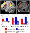
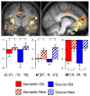
Post-retrieval processes are engaged when the outcome of a retrieval attempt must be monitored or evaluated. Functional neuroimaging studies have implicated right dorsolateral prefrontal cortex (DLPFC) as playing a role in post-retrieval processing. The present study used fMRI to investigate whether retrieval-related neural activity in DLPFC is associated specifically with monitoring the episodic content of a retrieval attempt. During study, subjects were cued to make one of two semantic judgments on serially presented pictures. One study phase was followed by a source memory task, in which subjects responded 'new' to unstudied pictures, and signaled the semantic judgment made on each studied picture. A separate study phase was followed by a task in which the studied items were subjected to a judgment about their semantic attributes. Both tasks required that retrieved information be evaluated prior to response selection, but only the source memory task required evaluation of retrieved episodic information. In both tasks, activity in a common region of right DLPFC was greater for studied than for unstudied items, and the magnitude of this effect did not differ between the tasks. Together with the results of a parallel event-related potential study [Hayama, H. R., Johnson, J. D., & Rugg, M. D. (2008). The relationship between the right frontal old/new ERP effect and post-retrieval monitoring: Specific or non-specific? Neuropsychologia, 46(5), 1211-1223, doi:S0028-3932(07)00390-9], the present findings indicate that putative right DLPFC correlates of post-retrieval processing are not associated exclusively with monitoring or evaluating episodic content. Rather, the effects likely reflect processing associated with monitoring or decision-making in multiple cognitive domains.
Figures


Similar articles
-
Age-related changes in right middle frontal gyrus volume correlate with altered episodic retrieval activity.J Neurosci. 2011 Dec 7;31(49):17941-54. doi: 10.1523/JNEUROSCI.1690-11.2011. J Neurosci. 2011. PMID: 22159109 Free PMC article.
-
The relationship between the right frontal old/new ERP effect and post-retrieval monitoring: specific or non-specific?Neuropsychologia. 2008 Apr;46(5):1211-23. doi: 10.1016/j.neuropsychologia.2007.11.021. Epub 2007 Dec 4. Neuropsychologia. 2008. PMID: 18234241 Free PMC article.
-
Framing memories: How the retrieval query format shapes the neural bases of remembering.Neuropsychologia. 2016 Aug;89:309-319. doi: 10.1016/j.neuropsychologia.2016.06.036. Epub 2016 Jun 29. Neuropsychologia. 2016. PMID: 27373768
-
Prefrontal contributions to domain-general executive control processes during temporal context retrieval.Neuropsychologia. 2008 Mar 7;46(4):1088-103. doi: 10.1016/j.neuropsychologia.2007.10.023. Epub 2007 Nov 12. Neuropsychologia. 2008. PMID: 18155254 Free PMC article.
-
Memory search and the neural representation of context.Trends Cogn Sci. 2008 Jan;12(1):24-30. doi: 10.1016/j.tics.2007.10.010. Epub 2007 Dec 19. Trends Cogn Sci. 2008. PMID: 18069046 Free PMC article. Review.
Cited by
-
Decision Making and Fibromyalgia: A Systematic Review.Brain Sci. 2022 Oct 27;12(11):1452. doi: 10.3390/brainsci12111452. Brain Sci. 2022. PMID: 36358377 Free PMC article. Review.
-
Lack of effects of online HD-tDCS over the left or right DLPFC in an associative memory and metamemory monitoring task.PLoS One. 2024 Jun 7;19(6):e0300779. doi: 10.1371/journal.pone.0300779. eCollection 2024. PLoS One. 2024. PMID: 38848375 Free PMC article.
-
Age-related changes in right middle frontal gyrus volume correlate with altered episodic retrieval activity.J Neurosci. 2011 Dec 7;31(49):17941-54. doi: 10.1523/JNEUROSCI.1690-11.2011. J Neurosci. 2011. PMID: 22159109 Free PMC article.
-
Ventral fronto-temporal pathway supporting cognitive control of episodic memory retrieval.Cereb Cortex. 2015 Apr;25(4):1004-19. doi: 10.1093/cercor/bht291. Epub 2013 Oct 31. Cereb Cortex. 2015. PMID: 24177990 Free PMC article.
-
Decreased fronto-temporal interaction during fixation after memory retrieval.PLoS One. 2014 Oct 23;9(10):e110798. doi: 10.1371/journal.pone.0110798. eCollection 2014. PLoS One. 2014. PMID: 25340398 Free PMC article.
References
-
- Aggleton JP, Brown MW. Interleaving brain systems for episodic and recognition memory. Trends in Cognitive Sciences. 2006;10(10):455–63. doi:S1364–6613(06)00193-8. - PubMed
-
- Andrade A, Paradis AL, Rouquette S, Poline JB. Ambiguous results in functional neuroimaging data analysis due to covariate correlation. NeuroImage. 1999;10(4):483–6. doi:10493904. - PubMed
-
- Badre D, Wagner AD. Left ventrolateral prefrontal cortex and the cognitive control of memory. Neuropsychologia. 2007;45(13):2883–901. doi:S0028–3932(07)00221-7. - PubMed
-
- Brett M, Anton JL, Valabregue R, Poline JB. Region of interest analysis using an SPM toolbox. NeuroImage. 2002;16:2.
-
- Buckner RL, Andrews-Hanna JR, Schacter DL. The brain’s default network: anatomy, function, and relevance to disease. Annals of the New York Academy of Sciences. 2008;1124:1–38. doi:1124/1/1. - PubMed
Publication types
MeSH terms
Substances
Grants and funding
LinkOut - more resources
Full Text Sources

