. 2009 Jun 19;284(25):17138-17146.
doi: 10.1074/jbc.M109.002568. Epub 2009 Apr 21.

Chaperones of F1-ATPase

Affiliations

Chaperones of F1-ATPase

Anthony Ludlam et al. J Biol Chem. .

Abstract

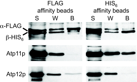
Mitochondrial F(1)-ATPase contains a hexamer of alternating alpha and beta subunits. The assembly of this structure requires two specialized chaperones, Atp11p and Atp12p, that bind transiently to beta and alpha. In the absence of Atp11p and Atp12p, the hexamer is not formed, and alpha and beta precipitate as large insoluble aggregates. An early model for the mechanism of chaperone-mediated F(1) assembly (Wang, Z. G., Sheluho, D., Gatti, D. L., and Ackerman, S. H. (2000) EMBO J. 19, 1486-1493) hypothesized that the chaperones themselves look very much like the alpha and beta subunits, and proposed an exchange of Atp11p for alpha and of Atp12p for beta; the driving force for the exchange was expected to be a higher affinity of alpha and beta for each other than for the respective chaperone partners. One important feature of this model was the prediction that as long as Atp11p is bound to beta and Atp12p is bound to alpha, the two F(1) subunits cannot interact at either the catalytic site or the noncatalytic site interface. Here we present the structures of Atp11p from Candida glabrata and Atp12p from Paracoccus denitrificans, and we show that some features of the Wang model are correct, namely that binding of the chaperones to alpha and beta prevents further interactions between these F(1) subunits. However, Atp11p and Atp12p do not resemble alpha or beta, and it is instead the F(1) gamma subunit that initiates the release of the chaperones from alpha and beta and their further assembly into the mature complex.


Figures

FIGURE 1.
FIGURE 1.
Pairwise alignment of recombinant protein sequences with S. cerevisiae homologs. Upper, pairwise alignment of C. glabrata and S. cerevisiae Atp11p. The disordered regions of the C. glabrata structure (residues 1–93 and 163–176) are shown in orange, the N-terminal helical domain (residues 94–129) in blue, the central α/β taco (residues 130–262) in green, and the C-terminal helical domain (residues 263–298) in red. Lower, pairwise alignment of P. denitrificans and S. cerevisiae Atp12p. The smaller N-terminal domain (residues 3–83) and the larger C-terminal domain (residues 84–238) are shown in blue and red, respectively. Trp-57 and Asp-202, corresponding, respectively, to Trp-103 and Glu-289 of the yeast protein, are highlighted in blue and red boxes. C. gla, C. glabrata; P. den, P. denitrificans; S. cer., S. cerevisiae.
FIGURE 2.
FIGURE 2.
Growth of S. cerevisiae mutants on nonfermentable and fermentable carbons. S. cerevisiae mutants disrupted at the genetic locus for Atp11p (Δatp11) or Atp12p (Δatp12) were transformed with a plasmid producing C. glabrata Atp11p or P. denitrificans Atp12p, and evaluated for growth on glucose and nonfermentable ethanol-glycerol (EG) plates. Wild type W303, the deletion strains Δatp11 and Δatp12, and the transformants Δatp11/YEp-cgatp11 and Δatp12/pRS316-pdatp12 were grown overnight in liquid YPD (2% glucose, 2% peptone, 1% yeast extract). The next day the cultures were adjusted to A600 = 1.0, and then serially diluted by a factor of 2. Five μl of each dilution were applied to a YPD and an EG plate (2% ethanol, 3% glycerol, 2% peptone, 1% yeast extract), and the plates were incubated at 30 °C. After 48 h, the deletion strain Δatp12 does not grow at all on EG, whereas Δatp11 displays a leaky phenotype. The transformant Δatp11/YEp-cgatp11, which produces from a multicopy plasmid C. glabrata Atp11p (56% identity with S. cerevisiae) with its own mitochondrial targeting sequence, grows on EG almost as well as the wild type. The transformant Δatp12/pRS316-pdatp12, which produces a chimeric protein in which P. denitrificans Atp12p (23% identity with S. cerevisiae) is fused to the mitochondrial targeting sequence of S. cerevisiae Atp11p, shows growth on EG, despite the plasmid being single copy; transformants of Δatp12 with the same chimeric allele expressed from a multicopy plasmid grow almost as well as the wild type (data not shown).
FIGURE 3.
FIGURE 3.
Structures of C. glabrata Atp11p and P. denitrificans Atp12p. A, N-terminal helical domain (residues 94–129), the central α/β taco (residues 130–262), and the C-terminal helical domain (residues 263–298) of C. glabrata Atp11p are shown as a cartoon colored in blue, green, and red, respectively. Loops are shown as lighter colors. B, solvent-accessible surface of Atp11p, rotated by ∼90° with respect to A, is colored in transparent light blue with the regions of identity with the S. cerevisiae protein shown in a darker shade; the internal structure is shown as a cartoon colored as in A. C, characteristic “boxing glove” shape of P. denitrificans Atp12p. The N-terminal “wrist” domain (residues 3–83), and the C-terminal “hand” domain (residues 84–238) are shown as a cartoon colored in shades of blue/light-blue and red/magenta, respectively. The first 37 residues of the P. denitrificans protein, corresponding to a deletion of the S. cerevisiae protein that severely affects stability, are shown in light blue. This region corresponds approximately to an N-terminal β-hairpin. The last 42 residues of the P. denitrificans protein, corresponding to a deletion of the S. cerevisiae protein that completely eliminates the chaperone activity, are shown in magenta. The surfaces of Trp-57 and Asp-202, corresponding to Trp-103 and Glu-289 of the yeast protein, are shown in yellow and green, respectively; mutations of these residues also inactivate the protein. D–F, palm (D) and dorso (E) faces of wild type Atp12p, and the palm face of D202K (F) Atp12p from P. denitrificans are shown colored according to the electrostatic potentials (−1 kT/e, red; 1 kT/e, blue) at their solvent-accessible surface. Figures were generated with PyMOL (36).
FIGURE 4.
FIGURE 4.
Details of the Atp12p structure. A, stereo view of the C-domain (from the top) showing a central helix surrounded by six other helices. B, relative motions of Atp12p N- and C-domains. Stereo view of four different structures of wild type Atp12p (shown as red, blue, yellow, and green ribbons) and of D202K Atp12p (shown as a cyan ribbon) superimposed through their N-domains to highlight the relative motion of the C-domain. C, detail of the 1.0 Å structure of wild type Atp12p around Asp-202 (visible in two conformations). D, detail of the 1.8 Å structure of D202K Atp12p around Lys-202. |2FoFc| σA maps are contoured at 1σ.
FIGURE 5.
FIGURE 5.
Western blots of sucrose gradient fractions to detect F1-ATPase assembly intermediates. Upper, purified yeast F1 (30 μg) or soluble extracts prepared by sonic irradiation of mitochondria (10 mg/ml) from wild type (WT) or Δγ isogenic yeast were mixed with molecular weight marker proteins (hemoglobin, 64 kDa; lipoamide dehydrogenase, 100 kDa) in 200 μl of buffer (20 mm Tris-HCl, pH 7.5, 4 mm ATP, 2 mm EDTA) and centrifuged through a 4.8-ml sucrose solution comprised of a linear 5–20% gradient (4.5 ml) formed over 80% sucrose (0.3 ml); the boundary between 20 and 80% sucrose is depicted by the vertical white dotted line in the schematic representation of the sucrose gradient at the top of the figure. The gradients were centrifuged in an SW55Ti rotor at 55,000 rpm for 2 h and 10 min at room temperature. Fractions (250 μl) collected from the bottom of the tubes were probed in Western blots using a mixture of polyclonal antibodies against α and β proteins and analyzed for the position of the marker proteins as described under “Experimental Procedures.” Arrowheads show the positions of hemoglobin (∼64 kDa), lipoamide dehydrogenase (∼100 kDa), and the F1 oligomer (∼360 kDa). Lower, to increase the resolution of F1 protein intermediate complexes observed in soluble extracts from Δγ yeast mitochondria (red dashed lines), the sample (100 μl) was centrifuged at room temperature through an expanded discontinuous gradient of sucrose (5–15% (4.6 ml) overlaying 80% (0.3 ml)) in an SW55Ti rotor at 28,200 rpm for 20 h. Fractions (250 μl) were analyzed in separate Western blots for the F1 α and β subunits, for Atp11p and for Atp12p. For ease of comparison, the three Western blots are shown here superimposed with their backgrounds digitally changed to three different colors as follows: yellow for α and β, green for Atp11p, and blue for Atp12p. The correspondence of peaks for Atp12p and F1 α, and for Atp11p and F1 β, is highlighted with the blue and green vertical lines, respectively.
FIGURE 6.
FIGURE 6.
Coprecipitation of proteins with F1 α-FLAG and with F1 β-HIS6. Western blots of proteins precipitated with affinity resins are shown (left, anti-FLAG M2 affinity gel (FLAG affinity beads); right, TALON metal affinity resin (HIS6 affinity beads)). Aliquots of the proteins present in the initial post-bead supernatants (S), in the first wash solutions (W), and in the collected bead fractions (B) were probed in separate Western blots using a mixture of polyclonal antibodies against F1 α and β or Atp11p or Atp12p antiserum. Total protein was precipitated with chloroform:MeOH from the supernatant and wash samples and suspended to the same volume of the recovered beads. Equivalent volumes (10 μl) of each fraction were loaded on the gels.
FIGURE 7.
FIGURE 7.
A possible interaction of Atp12p with F1 α. A, superposition of the yeast F1 γ subunit (cyan) to P. denitrificans Atp12p (yellow). The C-terminal helices of Atp12p are colored in red. B, predicted interaction of P. denitrificans Atp12p with yeast F1 α (green), based on the relative position of the α and γ subunits in yeast F1. (PDB code 2hld (Mol 1)).

Similar articles

Cited by

References

    1. Abrahams J. P., Leslie A. G., Lutter R., Walker J. E. (1994) Nature 370, 621–628 - PubMed
    1. Bianchet M. A., Hullihen J., Pedersen P. L., Amzel L. M. (1998) Proc. Natl. Acad. Sci. U. S. A. 95, 11065–11070 - PMC - PubMed
    1. Kabaleeswaran V., Puri N., Walker J. E., Leslie A. G., Mueller D. M. (2006) EMBO J. 25, 5433–5442 - PMC - PubMed
    1. Ackerman S. H., Tzagoloff A. (1990) Proc. Natl. Acad. Sci. U. S. A. 87, 4986–4990 - PMC - PubMed
    1. Lefebvre-Legendre L., Salin B., Schaëffer J., Brèthes D., Dautant A., Ackerman S. H., di Rago J. P. (2005) J. Biol. Chem. 280, 18386–18392 - PubMed

Publication types

MeSH terms

LinkOut - more resources