. 2009 Jun 19;284(25):17030-17038.
doi: 10.1074/jbc.M109.010066. Epub 2009 Apr 21.

The urokinase plasminogen activator receptor promotes efferocytosis of apoptotic cells

Affiliations

The urokinase plasminogen activator receptor promotes efferocytosis of apoptotic cells

Veera D'mello et al. J Biol Chem. .

Abstract

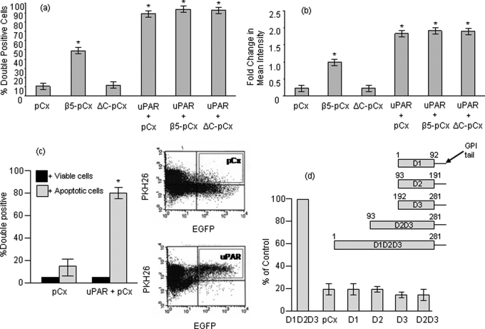
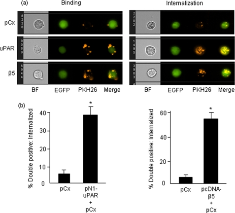
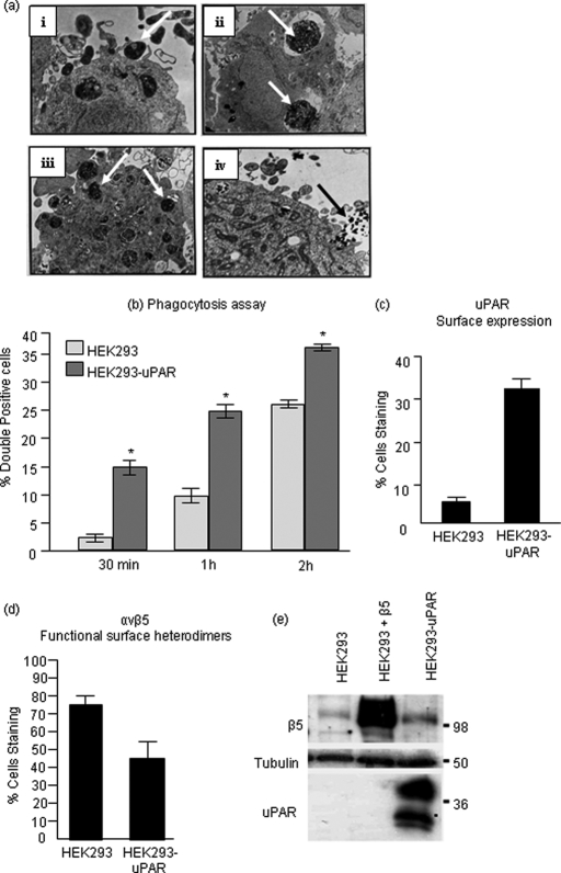
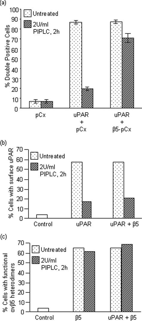
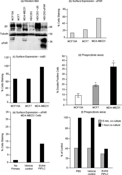
The urokinase receptor (uPAR), expressed on the surface of many cell types, coordinates plasmin-mediated cell surface proteolysis for matrix remodeling and promotes cell adhesion by acting as a binding protein for vitronectin. There is great clinical interest in uPAR in the cancer field as numerous reports have demonstrated that up-regulation of the uPA system is correlated with malignancy of various carcinomas. Using both stable cell lines overexpressing uPAR and transient gene transfer, here we provide evidence for a non-reported role of uPAR in the phagocytosis of apoptotic cells, a process that has recently been termed efferocytosis. When uPAR was expressed in human embryonic kidney cells, hamster melanoma cells, or breast cancer cells (BCCs), there was a robust enhancement in the efferocytosis of apoptotic cells. uPAR-expressing cells failed to stimulate engulfment of viable cells, suggesting that uPAR enhances recognition of one or more determinant on the surface of the apoptotic cell. uPAR-mediated engulfment was not inhibited by expression of mutant beta5 integrin, nor was alphavbeta5 integrin-mediated engulfment modulated by cleavage of uPAR by phosphatidylinositol-specific phospholipase C. Further, we found that the more aggressive BCCs had a higher phagocytic capacity that correlated with uPAR expression and cleavage of membrane-associated uPAR in MDA-MB231 BCCs significantly impaired phagocytic activity. Because efferocytosis is critical for the resolution of inflammation and production of anti-inflammatory cytokines, overexpression of uPAR in tumor cells may promote a tolerogenic microenvironment that favors tumor progression.


Figures

FIGURE 1.
FIGURE 1.
uPAR enhances phagocytosis independent of αvβ5 integrin. a, percentage of transfected green CS-1 cells that are positive for red-labeled apoptotic cells after 2 h of co-culture. CS-1 cells were transfected with pCx-EGFP or co-transfected with pCx-EGFP and pN1-uPAR or pN1-uPAR and β5-pCx or pN1-uPAR and the non-functional deletion mutant of β5 (ΔC-pCx). b, fold change in mean geometric intensity of the red channel (Y geometric mean) in the upper right quadrant that is representative of double positive population was measured. Values represent the amount of apoptotic material engulfed by phagocytes. Data are representative of at least three different experiments. c, uptake of viable or apoptotic T cells by EGFP and uPAR overexpressing cells after 2 h of co-culture. Co-culture of viable CEM-1 cells with transfected CS-1 cells resulted in ∼5% double-positive cells that correspond to basal cell death level in culture conditions in vitro. Panels on the right show graphical output of flow cytometry analysis showing engulfment represented in the upper right quadrant. d, the amino acid residues spanning each of the truncation mutants are depicted schematically. CS-1 cells were transfected with pCx-EGFP alone or together with the GPI-anchor containing pcDNA3-uPAR domain constructs as indicated, and a phagocytosis assay was performed. Values were normalized against D1D2D3, which was set at 100%. Asterisk, p < 0.05.
FIGURE 2.
FIGURE 2.
Analysis of phagocytosis by Amnis ImageStream System - Comparison of binding versus internalization. a, output images after Amnis acquisition. CS-1 cells were transiently transfected as shown in the figure and co-cultured with apoptotic cells for 2 h. Panels on the left depict peripheral binding of apoptotic components. Right panels were obtained after subtraction of two peripheral green pixels from phagocytes and depict internalized apoptotic material. BF, brightfield. b, quantification of internalized apoptotic material. Note that quantification does not include peripheral binding values and only represent phagocytes that have completely engulfed apoptotic bodies. Asterisk, p < 0.05.
FIGURE 3.
FIGURE 3.
Stable overexpression of uPAR in HEK293 cells accelerates corpse uptake. a, electron micrograph of HEK293 cell, a non-professional phagocyte engulfing apoptotic bodies indicated by arrowheads in respective panels. i, filopodial extensions and formation of phagocytic cup around an apoptotic body; ii and iii, presence of multiple apoptotic bodies within HEK293 cell; iv, a phagocytic portal engulfing multiple apoptotic blebs. b, HEK293 and HEK293-uPAR cells were labeled with membrane labeling dye PKH67 and co-cultured with the phagocytes at a ratio of 1:10. As cells were harvested by trypsinization, the above data represents mostly internalization. c, surface uPAR expression in HEK293-uPAR stable cells was confirmed by rabbit polyclonal (399R) antibody staining of live cells. d, cell surface expression of functional αvβ5 heterodimers in HEK293 and HEK293-uPAR cells was analyzed by monoclonal P1F6 antibody that recognizes a shared epitope in the heterodimer. e, Western blot analysis following extraction in SDS-containing radioimmune precipitation buffer. Asterisk, p < 0.05.
FIGURE 4.
FIGURE 4.
Deletion of cell-surface uPAR abrogates the observed phagocytic effect due to uPAR expression. a, CS-1 cells were transfected as indicated. The cells were either left untreated or treated with 2 units/ml glycophosphatidylinositol (GPI)-specific phospholipase C (PIPLC) that cleaves uPAR from the cell surface. Cells were then assessed for their phagocytic efficiency. b and c, surface expression of overexpressed proteins in transiently transfected CS-1 cells was confirmed by staining of live cells with primary antibodies followed by fluorescent secondary antibodies and analyzed by flow cytometry. Surface uPAR before and after PIPLC treatment was detected using a rabbit polyclonal 399R antibody (b). Functional αvβ5 heterodimers on the surface were confirmed by mouse monoclonal P1F6 antibody and were found to be intact after PIPLC treatment (c).
FIGURE 5.
FIGURE 5.
Relationship between uPAR expression and phagocytosis in BCCs. Two breast cancer cell lines (BCCs) viz. MCF7 and MDA-MB231 (in order of increasing invasiveness) and a normal cell line MCF10A were characterized for uPAR and β5 expression. a, protein expression was assessed by Western blotting. Note that in MDA-MB231 cells the major β5-integrin band runs as a faster migrating form. b and c, expression at the cell surface was analyzed by antibody staining of live cells followed by flow cytometric analysis. d, phagocytosis assay was performed by co-culturing MCF10A, MCF7, and MDA-MB231 cells with apoptotic T-cells for 2 h and analyzed by flow cytometry. e, MDA-MB231 cells were treated with vehicle or 8 units/ml PIPLC, and the reduction in the level of cell surface uPAR was assessed by antibody staining of live cells followed by flow cytometry. f, phagocytosis assay was carried out by co-culturing MDA-MB231 cells pre-treated with 8 units/ml PIPLC with apoptotic Jurkat cells for 15 min and 1 h in a phagocyte:apoptotic cell ratio of 1:5. Phagocytosis was compared with both PBS-treated and vehicle-treated cells. Asterisk, p < 0.05.

Similar articles

Cited by

References

    1. Roldan A. L., Cubellis M. V., Masucci M. T., Behrendt N., Lund L. R., Danø K., Appella E., Blasi F. ( 1990) EMBO J. 9, 467– 474 - PMC - PubMed
    1. Blasi F. ( 1993) Bioessays 15, 105– 111 - PubMed
    1. Rømer J., Bugge T. H., Pyke C., Lund L. R., Flick M. J., Degen J. L., Danø K. ( 1996) Nat. Med. 2, 725. - PubMed
    1. Romer J., Bugge T. H., Pyke C., Lund L. R., Flick M. J., Degen J. L., Dano K. ( 1996) Nat. Med. 2, 287– 292 - PubMed
    1. Saksela O., Rifkin D. B. ( 1988) Annu. Rev. Cell Biol. 4, 93– 126 - PubMed

MeSH terms

LinkOut - more resources