. 2009 Jul 15;18(14):2555-66.
doi: 10.1093/hmg/ddp187. Epub 2009 Apr 21.

Characterization of six human disease-associated inversion polymorphisms

Affiliations

Characterization of six human disease-associated inversion polymorphisms

Francesca Antonacci et al. Hum Mol Genet. .

Abstract

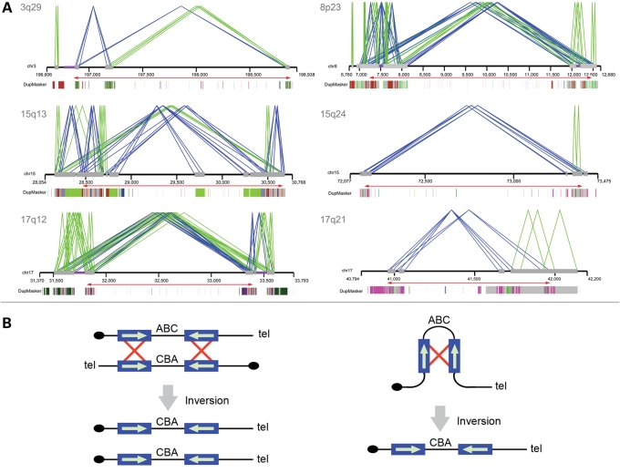
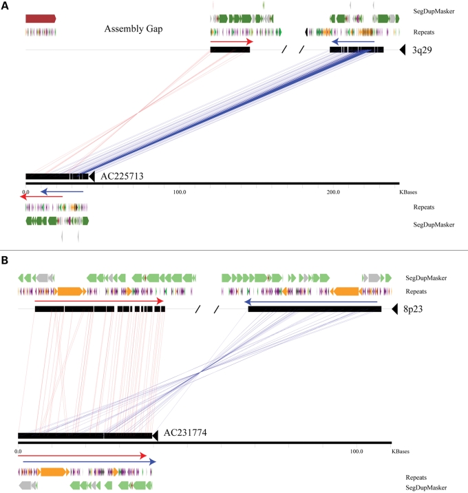
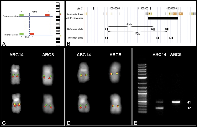
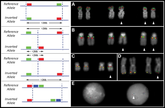
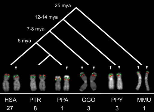
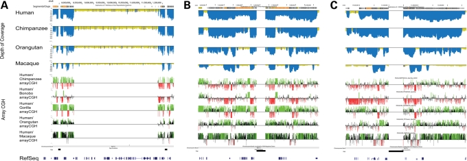
The human genome is a highly dynamic structure that shows a wide range of genetic polymorphic variation. Unlike other types of structural variation, little is known about inversion variants within normal individuals because such events are typically balanced and are difficult to detect and analyze by standard molecular approaches. Using sequence-based, cytogenetic and genotyping approaches, we characterized six large inversion polymorphisms that map to regions associated with genomic disorders with complex segmental duplications mapping at the breakpoints. We developed a metaphase FISH-based assay to genotype inversions and analyzed the chromosomes of 27 individuals from three HapMap populations. In this subset, we find that these inversions are less frequent or absent in Asians when compared with European and Yoruban populations. Analyzing multiple individuals from outgroup species of great apes, we show that most of these large inversion polymorphisms are specific to the human lineage with two exceptions, 17q21.31 and 8p23 inversions, which are found to be similarly polymorphic in other great ape species and where the inverted allele represents the ancestral state. Investigating linkage disequilibrium relationships with genotyped SNPs, we provide evidence that most of these inversions appear to have arisen on at least two different haplotype backgrounds. In these cases, discovery and genotyping methods based on SNPs may be confounded and molecular cytogenetics remains the only method to genotype these inversions.


Figures

Figure 1.
Figure 1.
Duplication architecture of inversion breakpoint regions. (A) Paralogy between large (≥10 kb), highly identical (≥95%) segmental duplications (gray bars) is shown. Direct (green lines) or inverted (blue lines) orientations of pairwise alignments of segmental duplications are indicated. A relaxed threshold (size ≥5 kbp and sequence identity ≥90%) was applied for the 15q24 inversion. The underlying ancestral duplication (duplicon) (24) composition of each duplication as determined by DupMasker (45) is represented as color-coded blocks. Red arrows show the extent of the inverted region. (B) Interchromosomal, intrachromosomal or intrachromatid non-allelic homologous recombination (NAHR) between inverted repeats causes inversion of the intervening sequence. Repeat sequences are indicated as blue boxes, with their orientation indicated by green arrows and recombination is shown by red crosses. Adapted in part from Sharp et al. (48).
Figure 2.
Figure 2.
Sequence resolution of inversion breakpoints. A miropeats comparison (49) between sequenced fosmids corresponding to the 3q29 (A) and 8p23 (B) inversions and the build35 reference are shown. For simplicity, only the regions around the breakpoints are depicted. Both inversion breakpoints map within highly identical stretches of duplicated sequence found in an inverted configuration (red and blue arrows), with the 3q29 inversion coincident with a gap in the reference assembly. Segmental duplications were predicted using DupMasker (45) and colored based upon the position of each ancestral duplicon. The locations of common repeats are also indicated: green: LINEs; purple: SINEs; orange: LTR elements; pink: DNA; gray/black: other/low complexity regions.
Figure 3.
Figure 3.
FISH inversion assay. (A) A schematic showing human genomic probes labeled in green and red mapping >2 Mb apart in the non-inverted state that appears as two distinct signals on chromosomal metaphase spreads. In the inverted state, the two probes map less the 2 Mb apart and appear as a merged yellow signal. Dashed blue lines indicate the inversion breakpoints. (B) We applied the FISH assay to distinguish the orientation of the 17q21.31 region on metaphase chromosomes. Human genomic fosmid probes A and B map >2 Mb apart in the non-inverted state and appear as two distinct signals (red and green) on chromosomal metaphase spreads. In contrast, probes A and B map less the 2 Mb apart in the inverted state and appear as a merged (red+green = yellow) signal (C). A reciprocal assay on the same samples using probes A and D (non-inverted=yellow; inverted=red+green) confirm the specificity of the assay (D). An analysis of 27 HapMap cell lines using this assay showed 100% correspondence between the H1/H2 haplotype and the non-inverted/inverted status as determined by PCR (E) and SNP genotyping.
Figure 4.
Figure 4.
FISH genotyping of inversion polymorphisms. (A) Metaphase FISH validation of the 8p23 inversion using two probes located inside of the inversion. Metaphase FISH-based assay to resolve inversions <2 Mb using one probe located inside and one outside the inverted region is shown for the 15q13.3 (B), 17q12 (C) and 3q29 (D) inversions. Interphase triple color FISH validation was used for the 15q24 inversion (E). Arrows indicate inverted chromosomes.
Figure 5.
Figure 5.
Comparative analysis of the 8p23 inversion. FISH validation of 8p23 inversion in 27 HapMap individuals, eight chimpanzees, one bonobo, three gorillas, three orangutans and one macaque showed the inverted orientation (H2) as the most likely ancestral state. The inversion polymorphism might have occurred in the shared human and chimpanzee lineage and still be polymorphic in human and bonobo, but fixed in the chimpanzee population. Arrows indicate inverted chromosomes.
Figure 6.
Figure 6.
(A) Comparative segmental duplication analysis of the 8p23 inversion region. The top panel shows the computationally predicted regions of segmental duplications [excess depth of coverage (blue) of aligned human, chimpanzee, orangutan and macaque WGS sequence to the human reference genome (build35)]. The lower panel shows the results of arrayCGH experiments for chimpanzee, bonobo, gorilla, orangutan and macaque using human as test. Enlargement of the distal and proximal breakpoints are shown in (B) and (C), respectively.

Similar articles

Cited by

References

    1. Iafrate A.J., Feuk L., Rivera M.N., Listewnik M.L., Donahoe P.K., Qi Y., Scherer S.W., Lee C. Detection of large-scale variation in the human genome. Nat. Genet. 2004;36:949–951. - PubMed
    1. Sebat J., Lakshmi B., Troge J., Alexander J., Young J., Lundin P., Maner S., Massa H., Walker M., Chi M., et al. Large-scale copy number polymorphism in the human genome. Science. 2004;305:525–528. - PubMed
    1. Sharp A.J., Locke D.P., McGrath S.D., Cheng Z., Bailey J.A., Vallente R.U., Pertz L.M., Clark R.A., Schwartz S., Segraves R., et al. Segmental duplications and copy-number variation in the human genome. Am. J. Hum. Genet. 2005;77:78–88. - PMC - PubMed
    1. Wong K.K., deLeeuw R.J., Dosanjh N.S., Kimm L.R., Cheng Z., Horsman D.E., MacAulay C., Ng R.T., Brown C.J., Eichler E.E., et al. A comprehensive analysis of common copy-number variations in the human genome. Am. J. Hum. Genet. 2007;80:91–104. - PMC - PubMed
    1. Perry G.H., Ben-Dor A., Tsalenko A., Sampas N., Rodriguez-Revenga L., Tran C.W., Scheffer A., Steinfeld I., Tsang P., Yamada N.A., et al. The fine-scale and complex architecture of human copy-number variation. Am. J. Hum. Genet. 2008;82:685–695. - PMC - PubMed

Publication types