. 2009 Jun;15(6):1059-66.
doi: 10.1261/rna.1606309. Epub 2009 Apr 21.

The C-terminal domains of human TNRC6A, TNRC6B, and TNRC6C silence bound transcripts independently of Argonaute proteins

Affiliations

The C-terminal domains of human TNRC6A, TNRC6B, and TNRC6C silence bound transcripts independently of Argonaute proteins

Daniela Lazzaretti et al. RNA. 2009 Jun.

Abstract

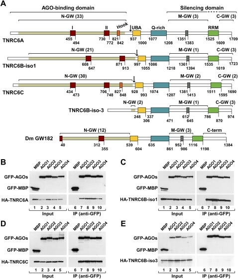
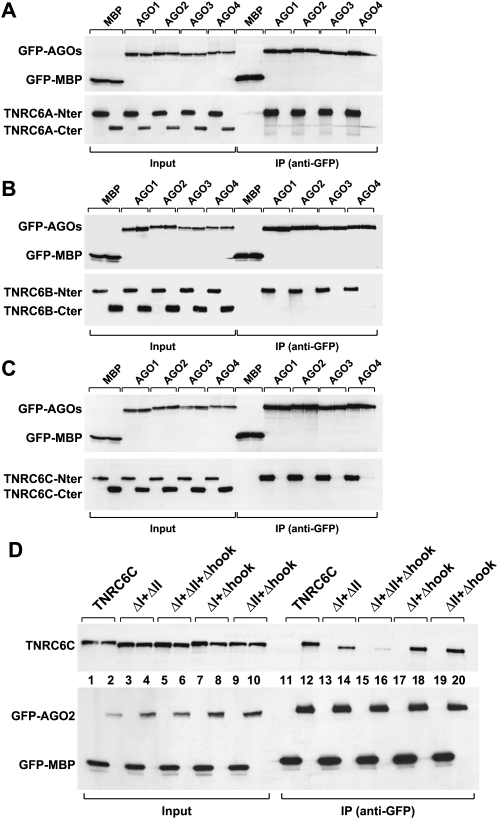
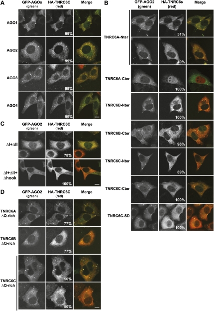
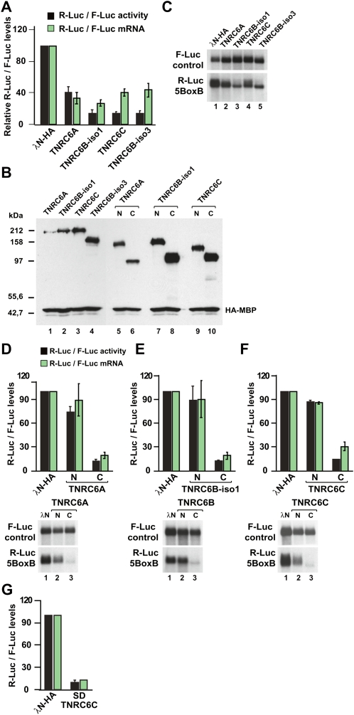
Proteins of the GW182 family are essential components of the miRNA pathway in animal cells. Vertebrate genomes encode three GW182 paralogs (TNRC6A, TNRC6B, and TNRC6C), which may be functionally redundant. Here, we show that the N-terminal GW-repeat-containing regions of all three TNRC6s interact with the four human Argonaute proteins (AGO1-AGO4). We also show that TNRC6A, TNRC6B, and TNRC6C silence the expression of bound mRNAs. This activity is mediated by their C-terminal silencing domains, and thus, is independent of the interaction with AGO1-AGO4. Silencing by TNRC6A, TNRC6B, and TNRC6C is effected by changes in protein expression and mRNA stability that can, in part, be attributed to deadenylation. Our findings indicate that TNRC6A, TNRC6B, and TNRC6C are recruited to miRNA targets through an interaction between their N-terminal domain and an Argonaute protein; the TNRC6s then promote translational repression and/or degradation of miRNA targets through a C-terminal silencing domain.


Figures

FIGURE 1.
FIGURE 1.
TNRC6A, TNRC6B, and TNRC6C interact with human AGO1–AGO4. (A) Domain organization of human TNRC6A–TNRC6C and D. melanogaster GW182. N-GW, M-GW, and C-GW: N-terminal, middle, and C-terminal GW-repeat-containing regions, respectively (the number of GW-repeats for each region is indicated in brackets). The white sectors within the N-terminal regions do not contain GW-repeats. UBA (yellow box): ubiquitin associated-like domain; Q-rich (cyan box): region rich in glutamine; RRM (green box): RNA recognition motif. Red boxes I and II: two conserved motifs within the N-terminal GW repeats. (Orange box) Ago-hook motif; (gray box) conserved motif III in the middle region. Numbers underneath the protein outline represent amino acid positions at fragment boundaries for each protein. Black arrows indicate the boundaries of the N and C-terminal protein fragments used in this study. The protein domains sufficient for the interaction with AGO1 and for silencing are indicated. (B–E) GFP-AGO1–AGO4 and HA-TNRC6A–TNRC6C were coexpressed in human HEK293 cells as indicated. Cell lysates were immunoprecipitated using anti-GFP antibodies. GFP-MBP served as a negative control. Inputs (2%) and immunoprecipitates (30%) were analyzed by Western blotting using anti-GFP or anti-HA antibodies.
FIGURE 2.
FIGURE 2.
The N-terminal GW-repeat-containing regions of TNRC6A–TNRC6C are required for the interaction with AGO1–AGO4. (A–C) GFP-AGO1–AGO4 and HA-TNRC6A–TNRC6C N- and C-terminal fragments were coexpressed in human HEK293 cells as indicated. Cell lysates were immunoprecipitated using anti-GFP antibodies. GFP-MBP served as a negative control. Inputs and immunoprecipitates were analyzed by Western blot as described in Figure 1, B–E. (D) Wild-type HA-TNRC6C or mutants were coexpressed with GFP-AGO2 or GFP-MBP in human HEK293 cells as indicated. Cell lysates were immunoprecipitated using anti-GFP antibodies. Inputs (2%) and immunoprecipitates (50%) were analyzed as described in A–C.
FIGURE 3.
FIGURE 3.
The AGO-binding domains and the Q-rich regions are required for P-body localization. (A) Localization of HA-TNRC6C in HeLa cells expressing GFP-AGO1–AGO4. (B) Localization of HA-tagged N- or C-terminal fragments of HA-TNRC6A–TNRC6C in HeLa cells expressing GFP-AGO2. (C) Localization of TNRC6 mutants in HeLa cells expressing GFP-AGO2. (D) Localization of HA-TNRC6A, TNRC6B, and TNRC6C lacking the Q-rich regions in HeLa cells expressing GFP-AGO2. In all panels, the merged images show the GFP signal in green and the HA signal in red. The fraction of cells exhibiting a staining identical to that shown in the representative panel was determined by scoring at least 100 cells (showing Argonaute-containing foci) in two independent transfections performed per protein. Scale bar, 10 μm.
FIGURE 4.
FIGURE 4.
TNRC6A–TNRC6C silence the expression of bound transcripts independently of the Argonaute proteins. (A–C) Human HEK293 cells were transfected with a mixture of three plasmids: the R-Luc-5BoxB reporter or the R-Luc control lacking the BoxB sequences, a plasmid expressing firefly luciferase as a transfection control, and vectors expressing the λN-HA-peptide or λN-HA-TNRC6A–TNRC6C. Renilla luciferase activity and mRNA levels were normalized to those of the firefly luciferase and set to 100 in cells expressing the λN-HA-peptide alone. Mean values ± standard deviations from three to four independent experiments are shown for the R-Luc-5BoxB reporter. Data corresponding to the R-Luc control (lacking the BoxB sequences) are not shown. In B, the expression levels of the λN-HA-TNRC6A–TNRC6C proteins and fragments were analyzed by Western blot. HA-MBP served as a transfection control. C shows a representative Northern blot. (D–F) Human HEK293 cells were transfected with a mixture of three plasmids: the R-Luc-5BoxB reporter or the R-Luc control reporter lacking the BoxB sequences, a plasmid expressing firefly luciferase as a transfection control, and vectors expressing the λN-HA-peptide or N- and C-terminal fragments of λN-HA-TNRC6A–TNRC6C. Renilla luciferase activity and mRNA levels were normalized to those of the firefly luciferase and analyzed as described in A. Representative Northern blots are shown in the lower panels. (G) Silencing activity of TNRC6C silencing domain (SD) measured as described in A.

References

    1. Behm-Ansmant I., Rehwinkel J., Doerks T., Stark A., Bork P., Izaurralde E. mRNA degradation by miRNAs and GW182 requires both CCR4:NOT deadenylase and DCP1:DCP2 decapping complexes. Genes & Dev. 2006a;20:1885–1898. - PMC - PubMed
    1. Behm-Ansmant I., Rehwinkel J., Izaurralde E. MicroRNAs silence gene expression by repressing protein expression and/or by promoting mRNA decay. Cold Spring Harb. Symp. Quant. Biol. 2006b;71:523–530. - PubMed
    1. Ding L., Han M. GW182 family proteins are crucial for microRNA-mediated gene silencing. Trends Cell Biol. 2007;17:411–416. - PubMed
    1. Ding L., Spencer A., Morita K., Han M. The developmental timing regulator AIN-1 interacts with miRISCs and may target the argonaute protein ALG-1 to cytoplasmic P bodies in C. elegans . Mol. Cell. 2005;19:437–447. - PubMed
    1. El-Shami M., Pontier D., Lahmy S., Braun L., Picart C., Vega D., Hakimi M.A., Jacobsen S.E., Cooke R., Lagrange T. Reiterated WG/GW motifs form functionally and evolutionarily conserved ARGONAUTE-binding platforms in RNAi-related components. Genes & Dev. 2007;21:2539–2544. - PMC - PubMed

Publication types

MeSH terms