. 2009 Jun;15(6):1067-77.
doi: 10.1261/rna.1605509. Epub 2009 Apr 21.

A C-terminal silencing domain in GW182 is essential for miRNA function

Affiliations

A C-terminal silencing domain in GW182 is essential for miRNA function

Ana Eulalio et al. RNA. 2009 Jun.

Abstract

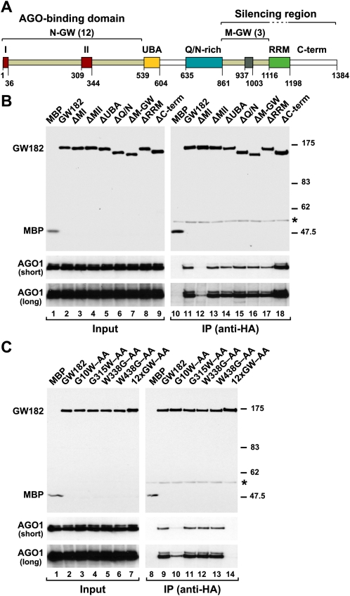
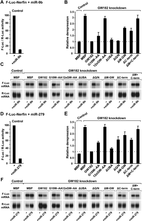
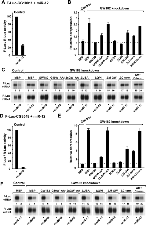
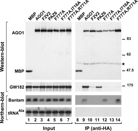
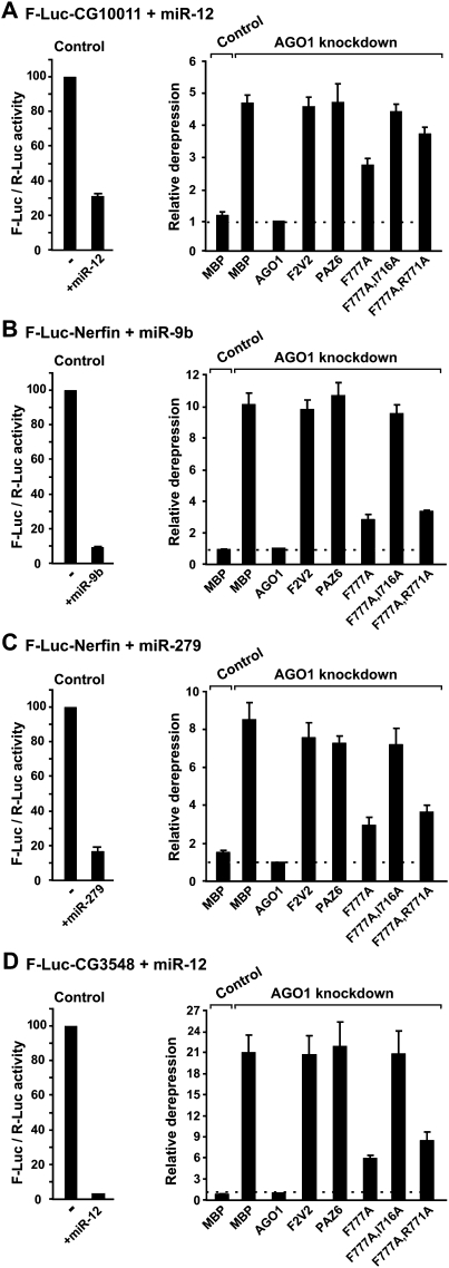
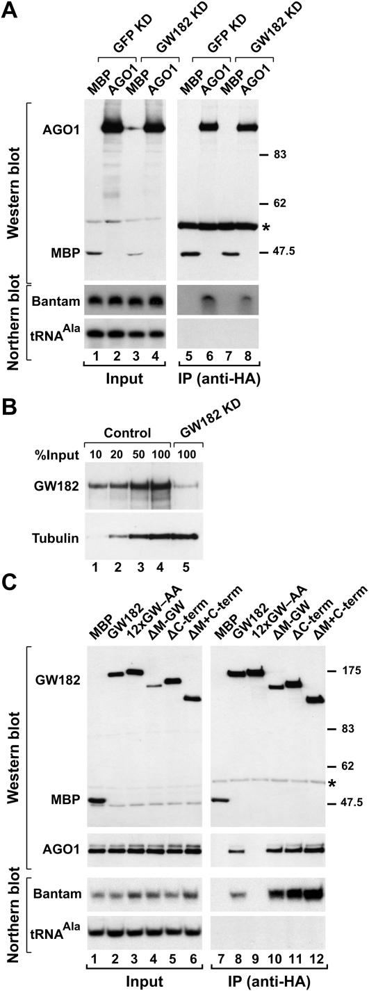
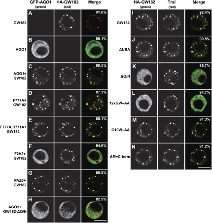
Proteins of the GW182 family are essential for miRNA-mediated gene silencing in animal cells; they interact with Argonaute proteins (AGOs) and are required for both the translational repression and mRNA degradation mediated by miRNAs. To gain insight into the role of the GW182-AGO1 interaction in silencing, we generated protein mutants that do not interact and tested them in complementation assays. We show that silencing of miRNA targets requires the N-terminal domain of GW182, which interacts with AGO1 through multiple glycine-tryptophan (GW)-repeats. Indeed, a GW182 mutant that does not interact with AGO1 cannot rescue silencing in cells depleted of endogenous GW182. Conversely, silencing is impaired by mutations in AGO1 that strongly reduce the interaction with GW182 but not with miRNAs. We further show that a GW182 mutant that does not localize to P-bodies but interacts with AGO1 rescues silencing in GW182-depleted cells, even though in these cells, AGO1 also fails to localize to P-bodies. Finally, we show that in addition to the N-terminal AGO1-binding domain, the middle and C-terminal regions of GW182 (referred to as the bipartite silencing domain) are essential for silencing. Together our results indicate that miRNA silencing in animal cells is mediated by AGO1 in complex with GW182, and that P-body localization is not required for silencing.


Figures

FIGURE 1.
FIGURE 1.
A GW-repeat in motif I of D. melanogaster GW182 provides a major binding site for AGO1. (A) Domain organization of GW182. N-GW and M-GW: N-terminal and middle GW-repeat-containing regions, respectively, with the number of repeats indicated in brackets; (UBA) ubiquitin associated-like domain; (Q/N-rich) region rich in glutamine (16.7%) and asparagine (14.5%); (RRM) RNA recognition motif; (C-term) C-terminal region. Red boxes I and II: two conserved motifs within the N-terminal GW-repeats. Gray box: conserved motif III in the middle region. The bipartite silencing region includes the M-GW and C-terminal regions but not the RRM, which is dispensable for silencing (Eulalio et al. 2009a). Amino acid positions at domain boundaries are indicated. (B,C) Lysates from S2 cells expressing HA-tagged versions of MBP, wild-type GW182, or GW182 mutants were immunoprecipitated using a monoclonal anti-HA antibody. Inputs (1.5%) and immunoprecipitates (30%) were analyzed by Western blotting using a polyclonal anti-HA antibody. Endogenous AGO1 was detected by Western blotting using anti-AGO1 antibodies (two different exposures are shown, short and long). (Asterisks) Indicate cross-reactivity with the immunoglobulin heavy chain by the secondary antibody (IP panels).
FIGURE 2.
FIGURE 2.
Role of GW182 domains in silencing. (A–F) S2 cells were treated with dsRNA targeting the 5′- and 3′-UTRs of GW182 mRNA. Control cells were treated with GFP dsRNA. These cells were subsequently transfected with a mixture of three plasmids: one expressing the F-Luc-Nerfin-1 reporter; another expressing miRNA primary transcripts (+miR-9b or miR-279) or the corresponding empty vector (−); and a third expressing Renilla luciferase (R-Luc). Plasmids (25 ng) encoding wild-type HA-GW182, HA-GW182 mutants, or HA-MBP were included in the transfection mixtures, as indicated. Firefly luciferase activities were normalized to those of the Renilla luciferase transfection control and set to 100 in cells transfected with the empty vector (i.e., in the absence of the miRNAs). (A,D) Normalized Firefly luciferase activities in the absence or presence of miRNAs in control cells (i.e., cells treated with GFP dsRNA and transfected with MBP). (B,E) Relative fold de-repression for each condition. Mean values ± standard deviations from three independent experiments are shown. (C,F) Northern blot analysis of representative RNA samples shown in B and E.
FIGURE 3.
FIGURE 3.
Role of GW182 domains in silencing. (A–F) S2 cells were treated with dsRNA targeting the 5′- and 3′-UTRs of GW182 mRNA. Control cells were treated with GFP dsRNA. These cells were subsequently transfected with a mixture of three plasmids: one expressing the indicated F-Luc reporters; another expressing miR-12 primary transcripts (+miR-12) or the corresponding empty vector (−); and a third expressing Renilla luciferase (R-Luc). Plasmids (25 ng) encoding wild-type HA-GW182, HA-GW182 mutants, or HA-MBP were included in the transfection mixtures, as indicated. Firefly luciferase activities were normalized to those of the Renilla luciferase transfection control and analyzed as described in Figure 2. (C,F) Northern blot analysis of representative RNA samples shown in B and E.
FIGURE 4.
FIGURE 4.
Mutations in AGO1 that uncouple miRNA and GW182 binding. (A) Lysates from S2 cells expressing HA-tagged versions of MBP, wild-type AGO1, or AGO1 mutants were immunoprecipitated using a monoclonal anti-HA antibody. Inputs (1.5%) and immunoprecipitates (30%) were analyzed by Western blotting using a polyclonal anti-HA antibody. Endogenous GW182 was detected using anti-GW182 antibodies. The association between HA-AGO1 and endogenous bantam was analyzed by Northern blotting. tRNAAla served as a loading control. (Asterisk) IgG cross-reactivity.
FIGURE 5.
FIGURE 5.
AGO1 mutants impaired in GW182-binding are also impaired in the complementation assay. (A–D) S2 cells were transfected with (Control) GFP-siRNA or (AGO1 knockdown) AGO1-siRNA plus a mixture of three plasmids: one expressing the indicated F-Luc reporters; another expressing miRNA primary transcripts (+miRNA) or the corresponding empty vector (−); and a third expressing Renilla luciferase (R-Luc). Plasmids encoding siRNA-resistant versions of wild-type or mutant HA-AGO1 proteins were included in the transfection mixtures, as indicated. Control cells were cotransfected with a plasmid encoding HA-MBP. Firefly luciferase activities were normalized to those of the Renilla luciferase transfection control and analyzed as described in Figure 2.
FIGURE 6.
FIGURE 6.
GW182 functions downstream from miRNA loading onto AGO1. (A) S2 cells were treated (Control) GFP or (GW182 KD) GW182 dsRNAs on days 0 and 4, and transfected on day 6 with plasmids expressing HA-MBP or HA-AGO1. Proteins were immunoprecipitated using a monoclonal anti-HA antibody. Inputs (1.5%) and immunoprecipitates (30%) were analyzed by Western blotting using a polyclonal anti-HA antibody. The association between HA-AGO1 and miRNAs was analyzed by Northern blotting. tRNAAla served as a loading control for the Northern blots. (B) The effectiveness of the GW182 depletion was analyzed by Western blotting using anti-GW182 antibodies. (Lanes 1–4) Dilutions of control cells (treated with GFP dsRNA) were loaded. Tubulin served as a loading control. (C) Lysates from S2 cells expressing HA-tagged versions of MBP, wild-type GW182, or GW182 mutants were immunoprecipitated using a monoclonal anti-HA antibody. Inputs (1.5%) and immunoprecipitates (30%) were analyzed by Northern blotting, as described in panel A. Endogenous AGO1 was detected by Western blotting using anti-AGO1 antibodies.
FIGURE 7.
FIGURE 7.
GW182 Q/N-rich region is required for P-body localization. (A–H) GFP-tagged AGO1 or mutants were expressed in S2 cells. In panels C–H, the effect of cotransfecting HA-GW182 or HA-GW182 mutants on the localization of AGO1 was examined. The merged images show the GFP signal in green, the HA signal in red. (I–N) Confocal fluorescent micrographs of fixed S2 cells expressing HA-tagged fusions of full-length GW182 or GW182 mutants. Cells were stained with affinity-purified anti-Tral antibodies. The merged images show the HA signal in green and the anti-Tral signal in red. In all panels, the fraction of cells stained identically to the representative panel was determined by scoring at least 100 cells in two independent transfections performed per protein. Scale bar, 5 μm.

References

    1. Bagga S., Bracht J., Hunter S., Massirer K., Holtz J., Eachus R., Pasquinelli A.E. Regulation by let-7 and lin-4 miRNAs results in target mRNA degradation. Cell. 2005;122:553–563. - PubMed
    1. Behm-Ansmant I., Rehwinkel J., Doerks T., Stark A., Bork P., Izaurralde E. mRNA degradation by miRNAs and GW182 requires both CCR4:NOT deadenylase and DCP1:DCP2 decapping complexes. Genes & Dev. 2006a;20:1885–1898. - PMC - PubMed
    1. Behm-Ansmant I., Rehwinkel J., Izaurralde E. MicroRNAs silence gene expression by repressing protein expression and/or by promoting mRNA decay. Cold Spring Harb. Symp. Quant. Biol. 2006b;71:523–530. - PubMed
    1. Bhattacharyya S.N., Habermacher R., Martine U., Closs E.I., Filipowicz W. Relief of microRNA-mediated translational repression in human cells subjected to stress. Cell. 2006;125:1111–1124. - PubMed
    1. Chu C.Y., Rana T.M. Translation repression in human cells by microRNA-induced gene silencing requires RCK/p54. PLoS Biol. 2006;4:e210. doi: 10.1371/journal.pbio.0040210. - DOI - PMC - PubMed

Publication types