. 2009 May 5;106(18):7607-12.
doi: 10.1073/pnas.0900688106. Epub 2009 Apr 21.

Aberrant cleavage of TDP-43 enhances aggregation and cellular toxicity

Affiliations

Aberrant cleavage of TDP-43 enhances aggregation and cellular toxicity

Yong-Jie Zhang et al. Proc Natl Acad Sci U S A. .

Abstract

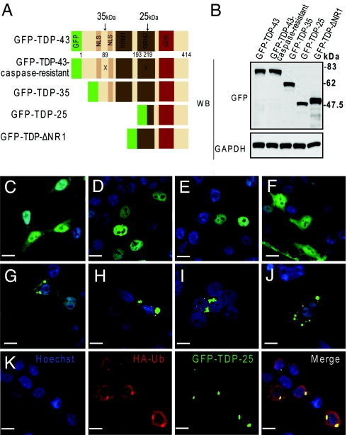
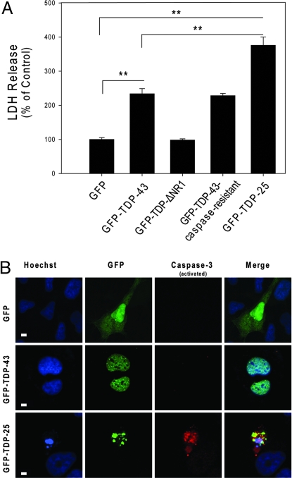
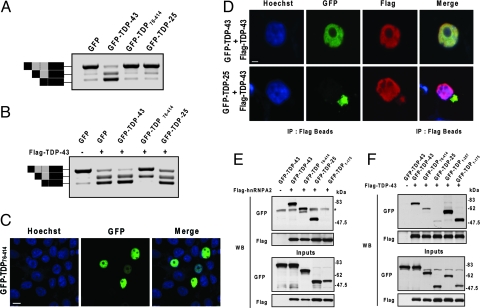
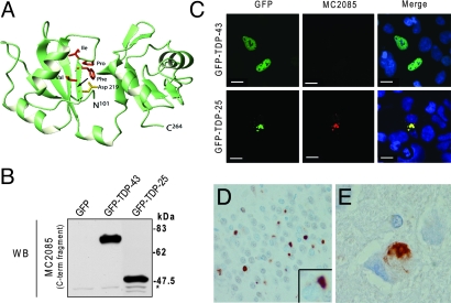
Inclusions of TAR DNA-binding protein-43 (TDP-43), a nuclear protein that regulates transcription and RNA splicing, are the defining histopathological feature of frontotemporal lobar degeneration with ubiquitin-positive inclusions (FTLD-Us) and sporadic and familial forms of amyotrophic lateral sclerosis (ALS). In ALS and FTLD-U, aggregated, ubiquitinated, and N-terminally truncated TDP-43 can be isolated from brain tissue rich in neuronal and glial cytoplasmic inclusions. The loss of TDP-43 function resulting from inappropriate cleavage, translocation from the nucleus, or its sequestration into inclusions could play important roles in neurodegeneration. However, it is not known whether TDP-43 fragments directly mediate toxicity and, more specifically, whether their abnormal aggregation is a cause or consequence of pathogenesis. We report that the ectopic expression of a approximately 25-kDa TDP-43 fragment corresponding to the C-terminal truncation product of caspase-cleaved TDP-43 leads to the formation of toxic, insoluble, and ubiquitin- and phospho-positive cytoplasmic inclusions within cells. The 25-kDa C-terminal fragment is more prone to phosphorylation at S409/S410 than full-length TDP-43, but phosphorylation at these sites is not required for inclusion formation or toxicity. Although this fragment shows no biological activity, its exogenous expression neither inhibits the function nor causes the sequestration of full-length nuclear TDP-43, suggesting that the 25-kDa fragment can induce cell death through a toxic gain-of-function. Finally, by generating a conformation-dependent antibody that detects C-terminal fragments, we show that this toxic cleavage product is specific for pathologic inclusions in human TDP-43 proteinopathies.


Conflict of interest statement

The authors declare no conflict of interest.

Figures

Fig. 1.
Fig. 1.
C-terminal, caspase-dependent fragments form cytoplasmic, ubiquitin-positive inclusions. (A) Schematic representation of the TDP-43 molecule and various TDP-43 constructs. The “X” symbol indicates 2 of the caspase-cleavage sites that result in either 35- or 25-kDa C-terminal TDP-43 fragments. NLS, nuclear localization signal; RRM, RNA recognition motif; GRR, glycine-rich region. (B) Expression of GFP fusion proteins in vitro. HEK293 cells were transfected with one of the constructs for 48 h, and cell lysates were subjected to Western blot analysis using an anti-GFP antibody to compare expression levels of the fusion proteins. The membrane was stripped and reprobed with an anti-GAPDH antibody to verify protein loading. (C–K) Fluorescent confocal microscopy demonstrates the diffuse cytoplasmic and nuclear distribution of GFP (C) and GFP-TDP-ΔNR1 (F), as well as the predominantly nuclear localization of wild-type GFP-TDP-43 (D) and the caspase-resistant mutant (E). Note the cytoplasmic inclusions formed in cells transfected with GFP-TDP-35 (G) or GFP-TDP-25 (H). Inclusions formed from the 25-kDa fragment persist after treatment with 0.2% Triton X-100 (I) or nocodazole (J). (K) Ubiquitin immunostaining shows that cytoplasmic inclusions formed from GFP-TDP-25 are ubiquitin-positive. (Scale bar: 10 μm.)
Fig. 2.
Fig. 2.
The 25-kDa C-terminal fragment of TDP-43 is prone to phosphorylation, but its phosphorylation is not required for inclusion formation. (A) Cells were transfected for GFP-TDP-43 or GFP-TDP-25, then immunostained with anti-pTDP-43, which detects TDP-43 when phosphorylated at S409/S410. Fluorescent confocal microscopy demonstrates enhanced staining of GFP-TDP-25 compared with GFP-TDP-43. (B) Lysates from HEK293 cells transfected with GFP-TDP-43, GFP-TDP-43NLSmut, GFP-TDP-25, or GFP-TDP-25S409A/S410A were subjected to Western blot analysis and probed with anti-pTDP-43. The 25-kDa C-terminal fragment showed marked anti-pTDP-43 immunoreactivity compared with GFP-TDP-43 or GFP-TDP-43NLSmut. GFP-TDP-25S409A/S410A did not exhibit any immunoreactivity. The membrane was reprobed with anti-GAPDH to verify protein loading. (C) Fluorescent confocal microscopy demonstrates that GFP-TDP-25S409A/S410A, which is not phosphorylated at S409/S410, can form cytoplasmic inclusions.
Fig. 3.
Fig. 3.
The 25-kDa C-terminal fragment enhances cellular toxicity. (A) The release of LDH into the media was used as an indicator of cell toxicity. LDH levels were measured 72 h after cells were transfected with the indicated constructs. Data from 3 separate experiments were analyzed by 1-way ANOVA, followed by Tukey's posthoc analysis (**, P < 0.001). (B) Increased apoptosis in differentiated M17 neuroblastoma cells expressing GFP-TDP-25 compared with cells expressing GFP alone or GFP-TDP-43. Cultures were fixed and stained with Hoechst to label nuclei (blue) and activated caspase-3 antibody (red). (Scale bar: 10 μM.)
Fig. 4.
Fig. 4.
The 25-kDa C-terminal fragment does not affect full-length TDP-43 function or cellular localization. (A and B) To examine the effect of C-terminal fragments on endogenous TDP-43 function, cells were cotransfected with a CFTR minigene construct and a vector encoding the GFP-fusion proteins, as indicated (A). To examine the effect of C-terminal fragments on exogenous TDP-43, cells were also transfected with a vector for FLAG-TDP-43 (B). Two days after transfection, the exon inclusion and exclusion products were examined by RT-PCR. The schematic shown to the left of the gels in A and B depicts transcripts containing or lacking exon 9 with or without a cryptic splice variant (gray box). Black boxes depict non-CFTR exons. Top and lower bands correspond to transcripts that include or exclude exon 9, respectively. The middle band represents the cryptic splice varient. Note that TDP-25 inhibited neither endogenous nor exogenous TDP-43 exon exclusion activity. Yet, GFP-TDP76–414 did markedly attenuate exogenous TDP-43 activity, despite its nuclear localization (C), which was examined by staining cells with the nuclear marker, Hoechst (blue), after transfection. (Scale bar: 10 μM.) (D) Fluorescent confocal microscopy reveals that GFP-TDP-25 inclusions fail to sequester wild-type FLAG-TDP-43. HEK293 cells were cotransfected with FLAG-TDP-43 and GFP-TDP-43 or GFP-TDP-25 and stained with Hoescht (blue) and anti-FLAG antibody (red). (Scale bar: 10 μM.) (E and F) Coimmunoprecipitation experiments reveal that GFP-TDP-25 and GFP-TDP76–414 bind strongly to hnRNPA2 (E) but only weakly to wild-type FLAG-TDP-43 (F). Note that GFP-TDP-43 and the C-terminal deletion products, GFP-TDP1–257 and GFP-TDP1–175, do bind FLAG-TDP-43, indicating that TDP-43 molecules interact and that their binding to one another is enhanced by the presence of the N terminal. (* represents a nonspecific band.)
Fig. 5.
Fig. 5.
A conformation-dependent TDP-43 antibody that detects C-terminal fragments is specific for pathologic inclusions in human TDP-43 proteinopathies. (A) Three-dimensional modeling of wild-type TDP-43 (amino acids 101–264). The positions of the 8 amino acid residues following the caspase cleavage motif, selected to generate a polyclonal antibody, are indicated. Note that aspartate 219, which is part of the caspase cleavage motif, is buried within the molecule (arrow). (B) Western blot analysis of lysates from HEK293 cells transfected with GFP only, GFP-TDP-43, or GFP-TDP-25 and probed with MC2085. *, Endogenous TDP-43. (C) MC2085 immunostaining of HEK293 cells transfected with GFP-TDP-43 or GFP-TDP-25. Note the absence of staining in cells expressing wild-type TDP-43 compared with those expressing the 25-kDa C-terminal fragment. (Scale bars: 10 μm.) (D and E) Immunostaining of tissue from FTLD-U brain (D) and ALS brain (E) with MC2085 shows cytoplasmic inclusions with little or no nuclear TDP-43. (Magnification: 40×.)

References

    1. Neumann M, et al. Ubiquitinated TDP-43 in frontotemporal lobar degeneration and amyotrophic lateral sclerosis. Science. 2006;314:130–133. - PubMed
    1. Arai T, et al. TDP-43 is a component of ubiquitin-positive tau-negative inclusions in frontotemporal lobar degeneration and amyotrophic lateral sclerosis. Biochem Biophys Res Commun. 2006;351:602–611. - PubMed
    1. Dreyfuss G, Kim VN, Kataoka N. Messenger-RNA-binding proteins and the messages they carry. Nat Rev Mol Cell Biol. 2002;3:195–205. - PubMed
    1. Krecic AM, Swanson MS. hnRNP complexes: Composition, structure, and function. Curr Opin Cell Biol. 1999;11:363–371. - PubMed
    1. Buratti E, et al. TDP-43 binds heterogeneous nuclear ribonucleoprotein A/B through its C-terminal tail: An important region for the inhibition of cystic fibrosis transmembrane conductance regulator exon 9 splicing. J Biol Chem. 2005;280:37572–37584. - PubMed

Publication types