. 2009 May 5;106(18):7420-5.
doi: 10.1073/pnas.0903033106. Epub 2009 Apr 21.

ERalpha as ligand-independent activator of CDH-1 regulates determination and maintenance of epithelial morphology in breast cancer cells

Affiliations

ERalpha as ligand-independent activator of CDH-1 regulates determination and maintenance of epithelial morphology in breast cancer cells

Maria Dafne Cardamone et al. Proc Natl Acad Sci U S A. .

Abstract

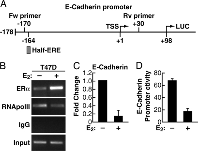
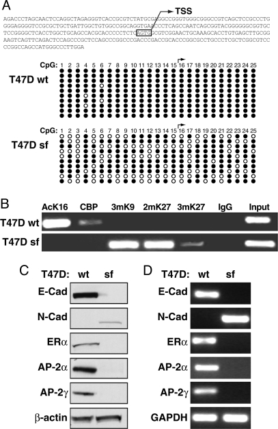
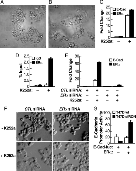
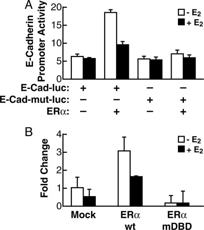
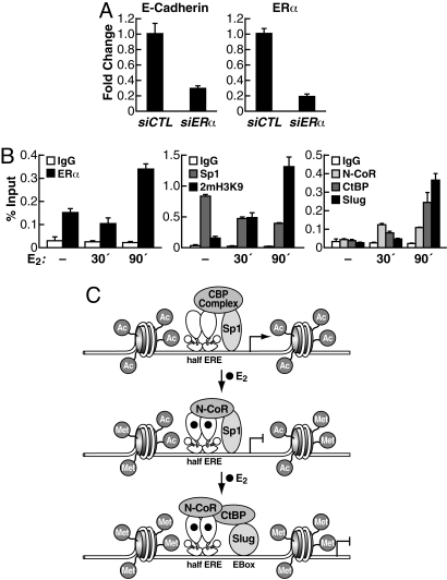
Estrogen receptor alpha (ERalpha) and E-cadherin are primary markers of luminal epithelial breast cancer cells with E-cadherin being a main caretaker of the epithelial phenotype. E-cadherin repression is needed for cancer cells to acquire motile and invasive properties, and it is known that in ER-positive breast cancer cells, estrogen down-regulate E-cadherin gene transcription. We report here that ERalpha is bound to the E-cadherin promoter in both the presence and the complete absence of estrogen, suggesting an unexpected role for unliganded ERalpha in E-cadherin transcription. Indeed, our data reveal that activation by unliganded ERalpha and repression by estrogen-activated ERalpha require direct binding to a half-estrogen response element within the E-cadherin promoter and exchange from associated coactivators to corepressors. Therefore, these results suggest a pivotal role for unliganded ERalpha in controlling a fundamental caretaker of the epithelial phenotype in breast cancer cells. Here, we show that ERalpha-positive breast cancer T47D cells transduced with the sfRON kinase undergo a full epithelial-mesenchymal conversion and lose E-cadherin and ERalpha expression. Our data show that, although the E-cadherin gene becomes hypermethylated and heterochromatic, kinase inhibitors can restore E-cadherin expression, together with an epithelial morphology in an ERalpha-dependent fashion. Similarly, transfection of ERalpha, in the absence of ligands, was sufficient to restore E-cadherin transcription in both sfRON-T47D and other ERalpha-, E-cadherin-negative cells. Therefore, our results suggest a novel role for the ERalpha that plays the dual role of ligand-independent activator and ligand-dependent repressor of E-cadherin in breast cancer cells.


Conflict of interest statement

The authors declare no conflict of interest.

Figures

Fig. 1.
Fig. 1.
Association of unliganded and ligand-activated ERα with E-cadherin promoter and effect on E-cadherin expression. (A) Schematic representation of the Luc-reporter construction, showing location of the half-site ERE at position −164/−160 in the E-cadherin promoter. (B) The ER+ T47D cells were grown in estrogen-free medium for 72 h. Then, the cells were treated with either ethanol vehicle (−E2) or E2 for 90 min. ChIP assay was performed with anti-ERα and anti-RNApolII antibodies. Input DNA was used to normalize the results. (C) Quantitative real-time PCR was used to evaluate changes in E-cadherin mRNA level in T47D cells similarly grown and treated with either ethanol vehicle (−E2) or E2 for 90 min. (D) T47D cells were grown in estrogen-free medium for 24 h, then were transiently transfected with E-cadherin promoter-Luc vector. After 48 h, cells were treated with either ethanol vehicle (−E2) or E2 for 90 min, and luciferase activity was measured and normalized by using β-gal activity.
Fig. 2.
Fig. 2.
Effect of a constitutively-active tyrosine kinase (sfRON) on chromatin organization at the E-cadherin gene promoter and change of ERα status in sfRON-T47D cells. (A) CpG methylation analysis of the CDH-1 promoter. The scheme shows the region analyzed. For each CpG (numbered from 5′ to 3′) black dots are unmethylated and open dots are methylated CpG. (B) Heterochromatic markers are enriched at the E-cadherin promoter in sfRON-T47D. ChIP assay was performed by using antibodies against acetyl-lysine 16 histone H4 (AcK16), trimethyl-lysine 9 of histone H3 (3mK9), dimethyl-lysine 27 of histone H3 (2mK27), trimethyl-lysine 27 of histone H3 (3mK27), and CBP. (C) Whole-cell protein extracts were subjected to immunoblotting for E-cadherin (E-Cad), N-cadherin (N-Cad), ERα, AP-2α, AP-2γ, and β-actin, showing equal protein loading. (D) End-point PCR was used to evaluate changes in E-cadherin (E-Cad), N-cadherin (N-Cad), ERα, AP-2α, and AP-2γ mRNA level as compared with GAPDH control.
Fig. 3.
Fig. 3.
Unliganded ERα is sufficient to activate the basal expression of E-cadherin in sfRON-T47D. (A and B) Reversion of the morphological penotype in sfRON-T47D cells by a kinase inhibitor. Cells were grown in estrogen-free medium for 72 h and then treated with K252a (B) or control vehicle (A) for 48 h. Images were taken at 40× magnification. (C) Quantitative real-time PCR was used to evaluate changes in E-cadherin and ERα mRNA level in sfRON-T47D cells in the presence of K252a or control vehicle. (D) K252a treatment induces ERα recruitment at the E-cadherin promoter. ChIP with quantitative real-time PCR was performed by using antibodies against ERα. (E) Induction of E-cadherin mRNA by K252a depends on ERα. sfRON-T47D cells were transfected with a control siRNA or a siRNA against ERα, grown in estrogen-free medium for 72 h, and treated with K252a or control vehicle. (F) The same experiment as in E demonstrates that ERα is required for morphological reversal in the presence of K252a. The images were taken at 40× magnification. (G) ERα expression induces E-cadherin-Luc reporter activity. Cells were grown in estrogen-free medium for 24 h then were transiently transfected with E-cadherin promoter-Luc vector, ERα-expressing vector, or empty vector. After 48 h luciferase activity was measured and normalized to β-gal activity.
Fig. 4.
Fig. 4.
Unliganded ERα regulates E-cadherin expression in HeLa cells. (A) HeLa cells were grown in estrogen-free medium for 24 h then transiently transfected with E-cadherin promoter-Luc vector or E-cadherin promoter-Luc vector ERE-dead mutant, along with ERα-expressing vector or empty vector. After 48 h of transfection, the cells were treated with either ethanol vehicle (−E2) or E2 for 90 min, and luciferase activity was measured and normalized to β-gal activity. (B) HeLa cells were grown as above and transiently transfected with of ERα wt or ERα mutDBD-expressing vectors or empty vector. After 48 h the cells were treated with either ethanol vehicle (−E2) or E2 for 90 min. Quantitative real-time PCR was used to evaluate changes in E-cadherin mRNA.
Fig. 5.
Fig. 5.
Dual role of unliganded or estrogen-activated ERα on E-cadherin gene expression in ER+ cells. (A) ERα is needed for basal E-cadherin expression in epithelial cells. MCF7 cells were grown in estrogen-free medium for 24 h then were transiently transfected with siRNA against ERα or control siRNA. After 48 h quantitative real-time PCR was used to evaluate changes in E-cadherin and ERα mRNA. (B) Increased ERα binding accompanies cofactor exchange at E-cadherin promoter after estradiol treatment. MCF7 cells were grown in estrogen-free medium for 72 h; the cells were treated with either ethanol vehicle (−E2) or E2 for 30 and 90 min. ChIP with quantitative real-time PCR was performed by using antibodies against ERα, histone H3 dimethyl-lysine 9 (2mH3K9), Sp1, NCoR (rabbit serum), CtBP, and Slug. (C) Proposed model for regulation of E-cadherin gene expression mediated by unliganded and estrogen-activated ERα.

References

    1. Perou CM, et al. Molecular portraits of human breast tumors. Nature. 2000;406:747–752. - PubMed
    1. Sorlie T, et al. Gene expression patterns of breast carcinomas distinguish tumor subclasses with clinical implications. Proc Natl Acad Sci USA. 2001;98:10869–10874. - PMC - PubMed
    1. Sotiriou C, et al. Breast cancer classification and prognosis based on gene expression profiles from a population-based study. Proc Natl Acad Sci USA. 2003;100:10393–10398. - PMC - PubMed
    1. Wirapati P, et al. Meta-analysis of gene expression profiles in breast cancer: Toward a unified understanding of breast cancer subtyping and prognosis signatures. Breast Cancer Res. 2008;10:R65. - PMC - PubMed
    1. Huber MA, Kraut N, Beug H. Molecular requirements for epithelial-mesenchymal transition during tumor progression. Curr Opin Cell Biol. 2005;17:548–558. - PubMed

Publication types