. 2009 May 1;69(9):3819-27.
doi: 10.1158/0008-5472.CAN-08-4389. Epub 2009 Apr 21.

The steroid receptor coactivator-1 regulates twist expression and promotes breast cancer metastasis

Affiliations

The steroid receptor coactivator-1 regulates twist expression and promotes breast cancer metastasis

Li Qin et al. Cancer Res. .

Abstract

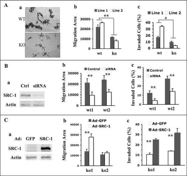
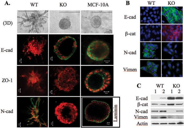
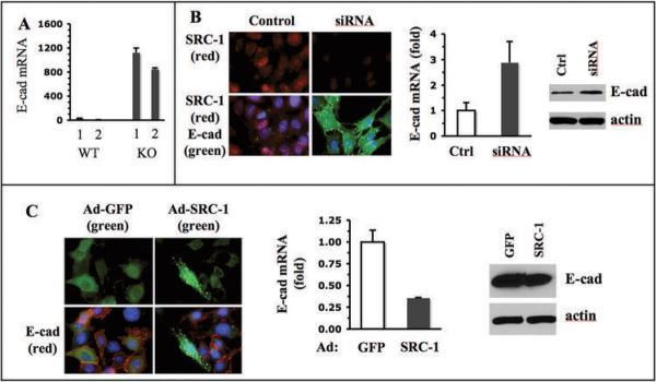
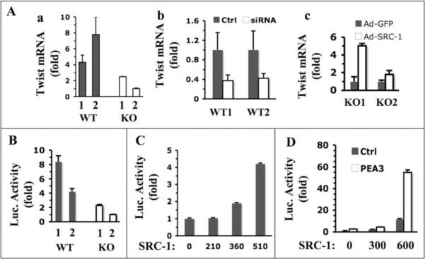
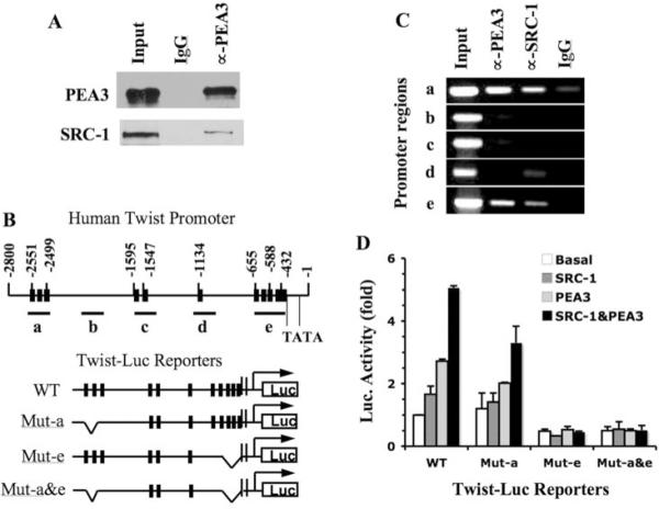
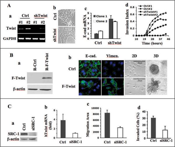
In breast cancer, steroid receptor coactivator-1 (SRC-1) expression positively correlates with HER2 expression and poor prognosis. In mouse mammary tumor virus-polyoma middle T (PyMT) breast cancer mouse model, SRC-1 strongly promotes mammary tumor metastasis. However, the molecular targets and mechanisms that mediate the role of SRC-1 in metastasis are unknown. In this study, SRC-1 wild-type (WT) and knockout (KO) cell lines were developed from the mammary tumors of WT/PyMT and KO/PyMT mice. WT cells exhibited strong migration and invasion capabilities, reduced E-cadherin and beta-catenin epithelial markers, gained N-cadherin and vimentin mesenchymal markers, and formed undifferentiated invasive structures in three-dimensional culture. In contrast, KO cells showed slow migration and invasion, retained E-cadherin, had less N-cadherin and vimentin, and developed partially differentiated three-dimensional structures. Importantly, WT cells expressed Twist, a master regulator of metastasis, at significantly higher levels versus KO cells. SRC-1 knockdown in WT cells reduced Twist expression, whereas SRC-1 restoration in KO cells also rescued Twist expression. Furthermore, SRC-1 was found to coactivate Twist transcription through physical interaction with the transcription factor PEA3 at the proximal Twist promoter. Accordingly, Twist knockdown in WT cells increased E-cadherin and reduced cell invasion and metastasis, and Twist expression in KO cells decreased E-cadherin and increased cell invasion. SRC-1 knockdown in human breast cancer cells also decreased Twist, cell migration, and invasion. Therefore, SRC-1 promotes breast cancer invasiveness and metastasis by coactivating PEA3-mediated Twist expression. Intervention of SRC-1 function may provide new strategies to inhibit breast cancer metastasis.


Figures

Fig. 1
Fig. 1. SRC-1 promotes mammary tumor cell migration and invasion
A. SRC-1 deficiency decreases tumor cell migration and invasion. The trails of WT and SRC-1 KO cell migration were traced using fluorescence beads (a). The migration areas of at least 40 cells were individually measured by pixels. The average migration areas per cell for WT1, WT2, KO1 and KO2 cell lines are presented (b). Cell migration data for these cell lines are presented as percentages of invaded cells to total cells in the invasion assay chambers (c). B. SRC-1 knockdown in WT cells decreases cell migration and invasion. Western blot analysis for SRC-1 and β-actin (loading control) in WT1 cells transfected with either scrambled double strand RNA (Ctrl) or SRC-1 siRNA (a). Cell migration and invasion data for WT1 and WT2 cells treated with control or SRC-1 siRNA are presented (b and c). C. SRC-1 restoration in SRC-1 KO cells rescues their migration and invasion. Western blot analysis confirmed SRC-1 expression in KO1 cells infected with SRC-1 adenoviruses. KO1 cells infected with GFP adenoviruses served as a control (a). The cell migration and invasion data for these cells are presented in panels b and c. *, p<0.05 and **, p<0.01 by unpaired t test.
Fig. 2
Fig. 2
SRC-1 deficiency helps to retain epithelial differentiation of mammary tumor cells. A. 3D structures formed from WT1, KO1 and MCF-10A cells. Immunofluorescence staining was performed for E-cadherin (E-cad), ZO-1, N-cadherin (N-cad) and Laminin. N-cad was not detected in the 3D structures of MCF-10A cells (data not shown). Laminin was not detected in the 3D structure of WT and KO mammary tumor cells (data not shown). B. Immunofluorescence staining for E-cad, β-catenin (β-cat), N-cad and vimentin (Vimen) in WT and KO mammary tumor cells cultured as monolayer. C. Western blot analyses of E-cad, β-cat, N-cad and Vimen in WT1, WT2, KO1 and KO2 mammary tumor cells. The β-actin served as a loading control.
Fig. 3
Fig. 3
SRC-1 inhibits E-cadherin expression. A. qPCR analysis of E-cadherin mRNA in WT1, WT2, KO1 and KO2 mammary tumor cells. The relative E-cadherin mRNA levels were normalized to the 18 S RNA. B. SRC-1 knockdown and Immunofluorescence staining of SRC-1 and E-cadherin in WT1 cells transfected with control or SRC-1 siRNA. The E-cadherin mRNA and protein levels in these cells were measured by qPCR and Western blot, respectively. C. SRC-1 expression and Immunostaining of SRC-1 and E-cadherin in KO1 mammary tumor cells with adenovirus-mediated GFP or SRC-1 expression. GFP signal and E-cadherin immunoreactive signal were imaged by fluorescence microscopy. The E-cadherin mRNA and protein levels in these cells were measured by qPCR and Western blot, respectively.
Fig. 4
Fig. 4
SRC-1 enhances Twist expression. A. qPCR analyses of relative Twist mRNA levels in the following groups of cells: WT1, WT2, KO1 and KO2 cells (a); WT1 and WT2 cells transfected with control or SRC-1 siRNA (b); and KO1 and KO2 cells with adenovirus-mediated GFP or SRC-1 expression (c). B. Transfection assays of Twist-Luc promoter/reporter construct in WT1, WT2, KO1 and KO2 cells. C. SRC-1 enhances Twist promoter activity in a dosage-dependent manner. Hela cells were co-transfected with Twist-Luc and different amounts (ng) of SRC-1 expression plasmids as indicated. D. SRC-1 and PEA3 synergistically enhance Twist promoter activity. Hela cells were transfected with Twist-Luc plasmids, variable amounts of SRC-1 expression plasmids as indicated, and with PEA3 expression plasmids or its parent vector. In all of the above transfection assays, the reporter luciferase activities were normalized to β-galactosidase activity from a co-transfected constitutively active expression vector.
Fig. 5
Fig. 5
SRC-1 directly regulates PEA3-mediated Twist expression. A. Co-IP analysis. MDA-MB-231 cell lysate was subjected to Co-IP with PEA3 antibody. None immunized IgG was used as a negative control. Input represents 5% of lysate used for Co-IP. Western blot analyses were performed with PEA3 and SRC-1 antibodies. B. PEA3 binding sites in the Twist promoter/enhancer region and Twist-Luc promoter/reporter constructs. The twist promoter region (bp −1 – −2800) contains 11 PEA3-binding elements (black bars). Fragments a–e were chosen for PCR amplification in ChIP assays. The positions of two TATA boxes were indicated. Four Twist-Luc promoter/reporter constructs in the pGL3 vector are depicted, which are WT, Mut-a (with deletions of the three PEA3-binding sites in region a), Mut-e (with deletions of the five PEA3 binding sites in region e) and Mut-a&e (with deletions of the PEA3 binding sites in both regions a and e). C. ChIP assays using MDA-MB-231 cells. Input represents 3% of material used for ChIP analysis. ChIP analyses were performed using PEA3 and SRC-1 antibodies. None immunized IgG was used as a negative control. PCR reactions were performed with specific primer pairs to amplify fragments a–e depicted in Panel B. D. Transfection assays for wild type and mutant Twist promoter activities. Hela cells were transfected with wild type or mutant Twist-Luc promoter/reporter constructs depicted in panel B. For each construct, cells also were co-transfected with parent vector (basal), SRC-1 and/or PEA3 expression plasmids as indicated. Data from three repeat assays are presented as mean ± SD.
Fig. 6
Fig. 6
Twist mediates the role of SRC-1 in breast cancer cells. A. Effects of Twist knockdown in WT1 cells. RT-PCR analysis revealed that Twist mRNA in WT1 cells was efficiently knocked down by lentivirus-mediated expression of two different shRNAs (#1 and #2) against mouse Twist mRNA. Lentivirus expressing non-targeting shRNAs served as a control (Ctrl) (a). The images of WT1 cells expressing control or Twist shRNA were taken under a phase-contrast microscope (b). Relative E-cadherin mRNA levels in WT1 cells expressing control or Twist shRNAs were measured by qPCR (c). Cell invasion indices of these cells were monitored in real time as described in Materials and Methods (d). B. Effects of Twist restoration in KO1 cells. Flag-tagged human Twist (F-Twist) was expressed in KO1 cells by retrovirus-mediated expression. Empty retrovirus served as a control. The F-Twist protein was detected by Flag antibody (a). Immunofluorescent staining of E-cadherin and vimentin in control and F-Twist-expressing KO1 cells was carried out using E-cadherin and vimentin antibodies. Cell images of 2D cultures and morphologies of 3D structures formed from control and F-Twist-expressing cells were taken under a phase-contrast microscope. C. Effects of SRC-1 knockdown in MDA-MB-231 cells. Cells were transfected with control or SRC-1 siRNAs. SRC-1 was analyzed by Western blot (a). Relative human Twist (hTwist) mRNA in these cells was measured by qPCR (b). Cell migration was traced with fluorescence beads. Average area (pixels) swept by each cell were obtained from measurements of at least 40 cells in each group (c). Cell invasion assay chambers with a Matrigel layer were used to determine the percentages of invaded cells to total cells (d). The assays were done in triplicates.

References

    1. Welch DR, Steeg PS, Rinker-Schaeffer CW. Molecular biology of breast cancer metastasis. Genetic regulation of human breast carcinoma metastasis. Breast Cancer Res. 2000;2:408–416. - PMC - PubMed
    1. Kauffman EC, Robinson VL, Stadler WM, Sokoloff MH, Rinker-Schaeffer CW. Metastasis suppression: the evolving role of metastasis suppressor genes for regulating cancer cell growth at the secondary site. J Urol. 2003;169:1122–1133. - PubMed
    1. Yang J, Mani SA, Donaher JL, Ramaswamy S, Itzykson RA, Come C, Savagner P, Gitelman I, Richardson A, Weinberg RA. Twist, a master regulator of morphogenesis, plays an essential role in tumor metastasis. Cell. 2004;117:927–939. - PubMed
    1. Thiery JP. Epithelial-mesenchymal transitions in tumour progression. Nat Rev Cancer. 2002;2:442–454. - PubMed
    1. Moustafa AS, Nicolson GL. Breast cancer metastasis-associated genes: prognostic significance and therapeutic implications. Oncol Res. 1997;9:505–525. - PubMed

Publication types

MeSH terms