. 2009 May 1;69(9):4036-42.
doi: 10.1158/0008-5472.CAN-08-3913. Epub 2009 Apr 21.

Analysis of DNA copy number alterations in ovarian serous tumors identifies new molecular genetic changes in low-grade and high-grade carcinomas

Affiliations

Analysis of DNA copy number alterations in ovarian serous tumors identifies new molecular genetic changes in low-grade and high-grade carcinomas

Kuan-Ting Kuo et al. Cancer Res. .

Erratum in

  • Cancer Res. 2009 Jun 15;69(12):5267

Abstract

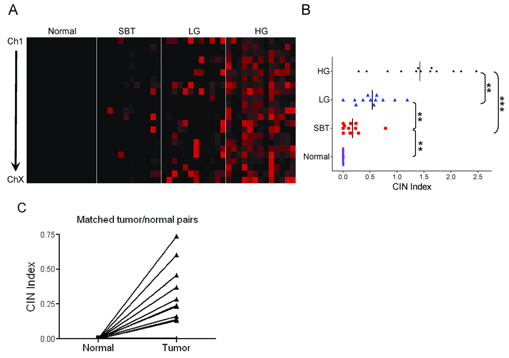
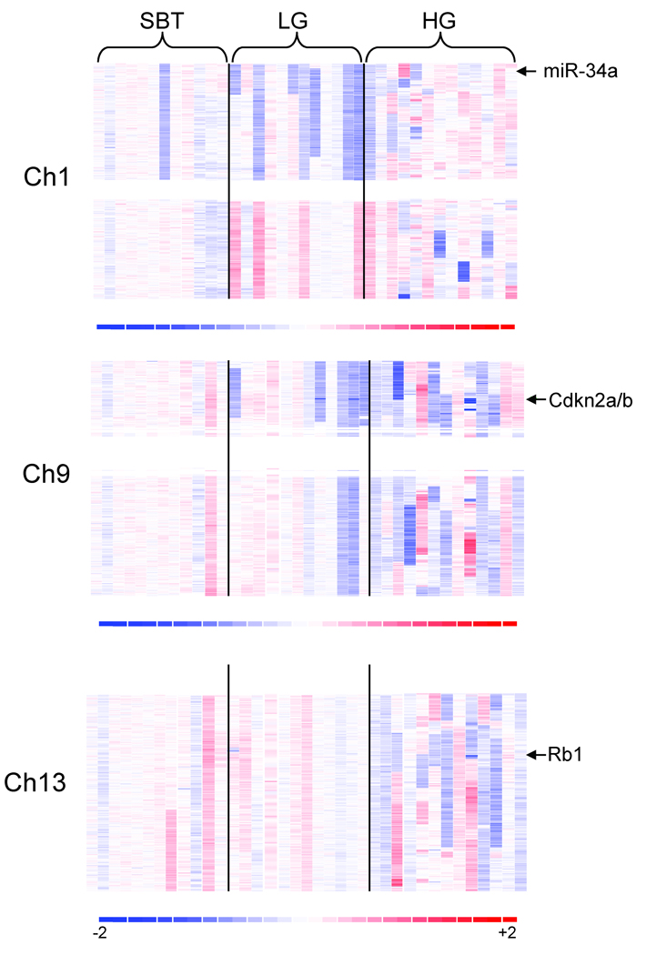
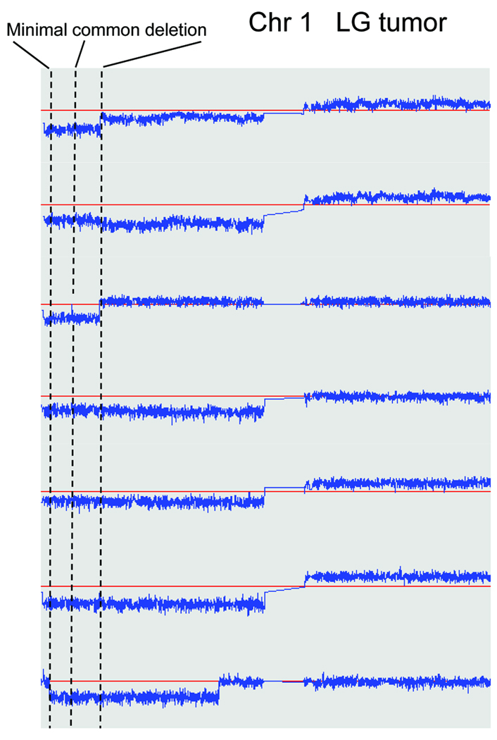
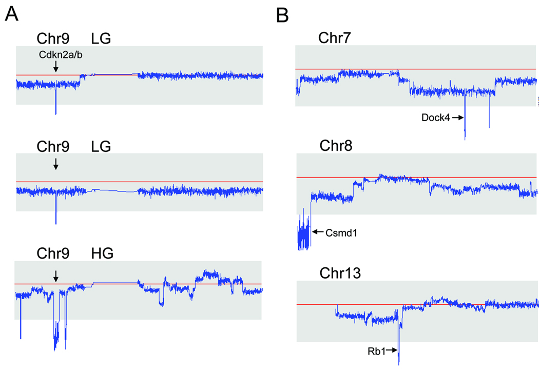
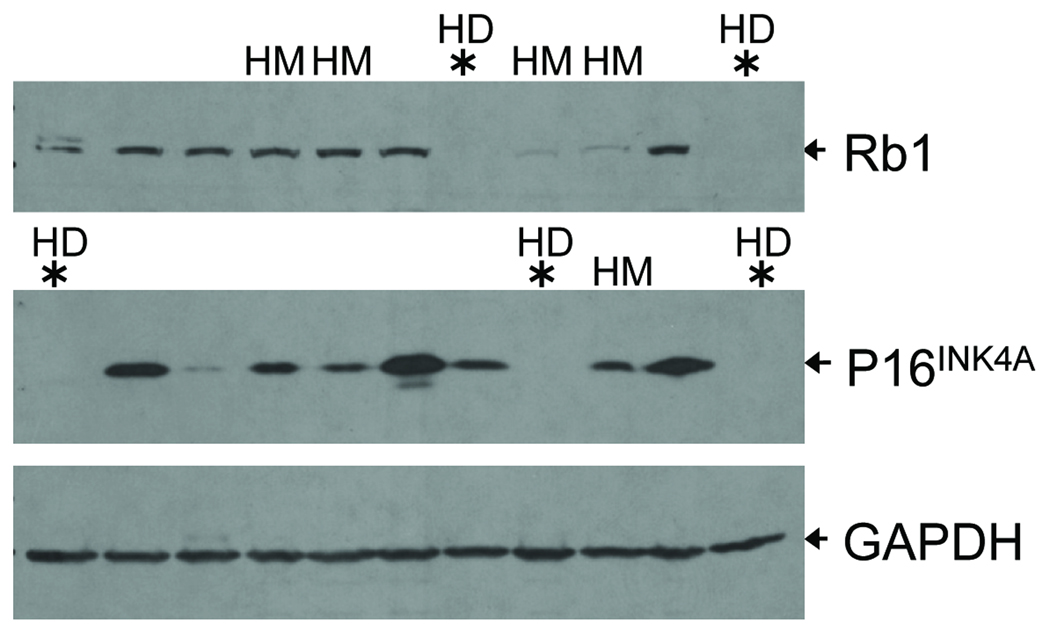
Ovarian serous carcinoma, the most common and lethal type of ovarian cancer, is thought to develop from two distinct molecular pathways. High-grade (HG) serous carcinomas contain frequent TP53 mutations, whereas low-grade (LG) carcinomas arise from serous borderline tumors (SBT) and harbor mutations in KRAS/BRAF/ERBB2 pathway. However, the molecular alterations involved in the progression from SBT to LG carcinoma remain unknown. In addition, the extent of deletion of tumor suppressors in ovarian serous carcinomas has not been well studied. To further address these two issues, we assessed DNA copy number changes among affinity-purified tumor cells from 37 ovarian serous neoplasms including SBT, LG, and HG tumors using high-density 250K single nucleotide polymorphism arrays. Chromosomal instability index as measured by changes in DNA copy number was significantly higher in HG than in LG serous carcinomas. Hemizygous ch1p36 deletion was common in LG serous carcinomas but was rarely seen in SBT. This region contains several candidate tumor suppressors including miR-34a. In contrast, in HG serous carcinomas, significant numbers of amplifications and deletions, including homozygous deletions, were identified. Among homozygous deletions, loci containing Rb1, CDKN2A/B, CSMD1, and DOCK4 were most common, being present in 10.6%, 6.4%, 6.4%, and 4.3%, respectively, in independent 47 affinity-purified HG serous carcinomas. Except for the CDKN2A/B region, these homozygous deletions were not present in either SBT or LG tumors. Our study provides a genome-wide homozygous deletion profile in HG serous carcinomas, which can serve as a molecular foundation to study tumor suppressors in ovarian cancer.


Figures

Figure 1
Figure 1. Genome-wide chromosome instability (CIN) index in ovarian serous tumors
A. The CIN index in individual chromosome of each serous tumor is plotted using a pseudo-color gradient indicating the copy number alteration level (low to high: dark to red). Normal: stromal fibroblast from the tumor; SBT: serous borderline tumor (atypical proliferative serous tumor); LG: low-grade serous carcinoma; HG: high-grade serous carcinoma. B. Genome-wide CIN index for each tumor. C. CIN index in matched normal and tumor pairs from 7 SBT and 6 LG tumors. Tumor samples are found to harbor significantly higher CIN index than adjacent normal samples (p<0.01).
Figure 2
Figure 2. Copy number alteration in chromosome 1, 9, and 13
DNA copy number changes are represented as pseudo-color gradients corresponding to the copy number increase (red boxes) and decrease (blue boxes) as compared to pooled normal samples. Each column represents an individual tumor sample. Arrow indicates candidate tumor suppressor gene at each region.
Figure 3
Figure 3. Minimal mapping of chromosome 1p hemizygous deletions in LG tumors
Copy number of chromosome 1 of all seven LG tumors harboring ch1p36 deletion is plotted against chromosomal location. The minimally deleted region (delineated by dotted lines) contains two adjacent deletion fragments. Red horizontal lines indicate normal copy number: 2. Candidate tumor suppressor genes, CHD5 and miRNA-34a, are located within the first deletion fragment.
Figure 4
Figure 4. Representative chromosomes containing homozygous deletions
A. Two LG serous carcinomas and one HG serous carcinoma harbor the ch9p21.3 homozygous deletion. Red horizontal lines represent normal copy number: 2. The deletions in LG serous carcinomas are small and discrete, but the deletion in the HG tumor is larger and encompasses 2.92 Mb. B. Representative homozygous deletions in ch7, ch8, and ch13 in HG serous carcinomas. Candidate tumor suppressor genes residing within the deleted regions are indicated by arrows.
Figure 5
Figure 5. Loss of p16 and Rb1 protein expression in tumors with homozygous deletion at 9p21.3 and 13q14.2, respectively
Western blot analysis was performed using HG serous tumors with known DNA copy number at 9p21.3 and 13q14.2 regions. Tumors with homozygous deletions (HD) at those loci were found to completely lose protein expression of p16 or Rb1, respectively. Hemizygous deletion (HM) was found to variably affect protein expression of p16 and Rb1. The remaining tumors contain normal copy number (2 copies). GAPDH was used as the sample loading control.
Figure 6
Figure 6. Biological effects of miR-34a in a LG carcinoma cell line, MPSC-1
(A) Quantitative PCR demonstrates a reduced genomic DNA (gDNA) copy number at the miR-34a locus in a LG carcinoma cell line, MPSC-1. The calculated copy number in normal tissue, MPSC-1, and 207T is 2.1, 0.8 and 1.0, respectively. 207T is a LG carcinoma with hemizygous deletion at the miR-34a locus based on SNP array analysis. (B) miR-34a expression measured by the TagMan microRNA assay. OSE: normal ovarian surface epithelial cells. (C) miR-34a inhibits cell proliferation as measured by the cell-titer blue assay. (D) mRNA levels of candidate miR-34a target genes measured by quantitative RT-PCR.

Similar articles

Cited by

References

    1. Jemal A, Siegel R, Ward E, et al. Cancer statistics, 2008. CA Cancer J Clin. 2008;58:71–96. - PubMed
    1. Link CJ, Jr, Kohn E, Reed E. The relationship between borderline ovarian tumors and epithelial ovarian carcinoma: epidemiologic, pathologic, and molecular aspects. Gynecol Oncol. 1996;60:347–354. - PubMed
    1. Singer G, Kurman RJ, Chang H-W, Cho SKR, Shih I-M. Diverse tumorigenic pathways in ovarian serous carcinoma. Am J Pathol. 2002;160:1223–1228. - PMC - PubMed
    1. Shih Ie M, Kurman RJ. Molecular pathogenesis of ovarian borderline tumors: new insights and old challenges. Clin Cancer Res. 2005;11:7273–7279. - PubMed
    1. Shih I-M, Kurman RJ. Ovarian tumorigenesis- a proposed model based on morphological and molecular genetic analysis. Am J Pathol. 2004;164:1511–1518. - PMC - PubMed

Publication types