. 2009 May-Jun;2(5-6):267-74.
doi: 10.1242/dmm.002261. Epub 2009 Apr 21.

FSHD region gene 1 (FRG1) is crucial for angiogenesis linking FRG1 to facioscapulohumeral muscular dystrophy-associated vasculopathy

Affiliations

FSHD region gene 1 (FRG1) is crucial for angiogenesis linking FRG1 to facioscapulohumeral muscular dystrophy-associated vasculopathy

Ryan D Wuebbles et al. Dis Model Mech. 2009 May-Jun.

Abstract

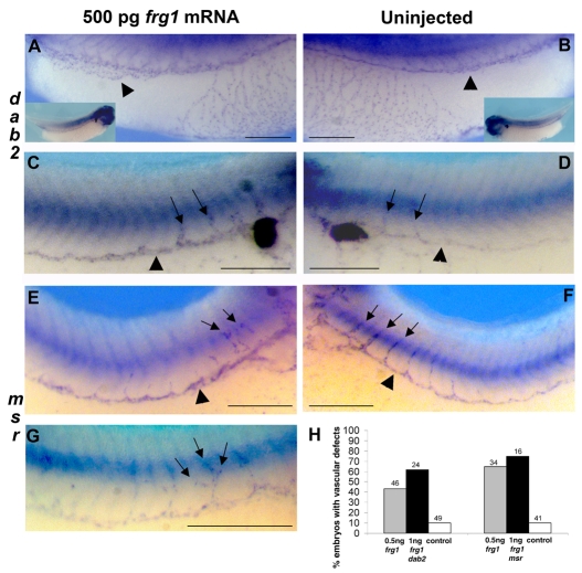
The genetic lesion that is diagnostic for facioscapulohumeral muscular dystrophy (FSHD) results in an epigenetic misregulation of gene expression, which ultimately leads to the disease pathology. FRG1 (FSHD region gene 1) is a leading candidate for a gene whose misexpression might lead to FSHD. Because FSHD pathology is most prominent in the musculature, most research and therapy efforts focus on muscle cells. Previously, using Xenopus development as a model, we showed that altering frg1 expression levels systemically leads to aberrant muscle development, illustrating the potential for aberrant FRG1 levels to disrupt the musculature. However, 50-75% of FSHD patients also exhibit retinal vasculopathy and FSHD muscles have increased levels of vascular- and endothelial-related FRG1 transcripts, illustrating an underlying vascular component to the disease. To date, no FSHD candidate gene has been proposed to affect the vasculature. Here, we focus on a role for FRG1 expression in the vasculature. We found that endogenous frg1 is expressed in both the developing and adult vasculature in Xenopus. Furthermore, expression of FRG1 was found to be essential for the development of the vasculature, as a knockdown of FRG1 resulted in decreased angiogenesis and reduced expression of the angiogenic regulator DAB2. Conversely, tadpoles subjected to frg1 overexpression displayed the pro-angiogenic phenotypes of increased blood vessel branching and dilation of blood vessels, and developed edemas, suggesting that their circulation was disrupted. Thus, the systemic upregulation of the FRG1 protein shows the potential for acquiring a disrupted vascular phenotype, providing the first link between a FSHD candidate gene and the vascular component of FSHD pathology. Overall, in conjunction with our previous analysis, we show that FRG1 overexpression is capable of disrupting both the musculature and vasculature, recapitulating the two most prominent features of FSHD.


Figures

Fig. 1.
Fig. 1.
Expression of the FRG1 protein during development. (A) Stage 36 whole-mount immunostaining shows the ubiquitous appearance of the FRG1 protein. FRG1-immunostained tadpoles were then sectioned either sagitally (B) or transversally (C) in the regions depicted in A. (D) Diagram of the minimal frg1 regulatory element (FRE) construct that was used to examine tissues with active transcription. EGFP expression was observed in stage 32 transgenic animals by both whole-mount fluorescence (E) and egfp in situ hybridization (F). By stage 36, transgenic egfp expression becomes restricted to muscle and vascular cell lineages (G), and by stage 42 it is observed almost exclusively within the vasculature (H). In adult X. laevis, immunostaining of FRG1 [green (I,K)] is observed in the gastrocnemius muscle, where it colocalizes precisely with rhodamine-labled lectin [red (J,K)], a marker for capillaries. Antibody specificity for the FRG1 peptide in these immunohistology experiments was confirmed by immunostaining with (M), or without (L), FRG1 peptide competition. (N) Strong FRG1 staining (red) was observed in arteries and veins in gastrocnemious sections co-stained with wheat germ agglutinin (green) and DAPI (blue). Abbreviations: ps, pronephric sinus; pd, pronephric duct; pcv, posterior cardinal vein; pda, paired dorsal arteries; nc, notochord; nt, neural tube; aa, aortic arches; ov, ophthalmic vessel; da, dorsal artery; dlav, dorsal lateral vein. Bars, 1 mm (A,E–H); 30 μm (B,C); 50 μm (K–N).
Fig. 2.
Fig. 2.
Depletion of FRG1 inhibits vascular development. The mild (A) and severe (B) phenotypes of the FMO1-injected (40 ng) embryos show a reduction in vascular structures compared with the uninjected sides of the same embryos (D,E), as visualized by dab2 expression. (C,F) CMO-injected and uninjected sides of the same embryo, respectively. (G,J) FMO1-injected (40 ng) and uninjected sides of the same embryo, stained for msr. The arrowheads point to intersomitic veins. (H,I) Partial and full rescue of dab2 staining by co-injection with 40 ng of FMO1 and 500 pg of X. tropicalis frg1 mRNA. Co-injection of 40 ng of FMO1 with 1 ng of X. tropicalis frg1 mRNA was lethal. (M) The percentage of injected embryos displaying loss of dab2 and msr staining. (N) The percentage of embryos rescued by co-injection of FMO1 with frg1 mRNA. The numbers above the bars indicate the total number of embryos analyzed. Abbreviations: ps, pronephric sinus; vvn, vitelline vein network; pcv, posterior cardinal vein. Bars, 0.5 mm.
Fig. 3.
Fig. 3.
Embryos with elevated FRG1 levels have increased angiogenesis and vascular dilation. (A–G) Stage 36 tadpoles injected with 500 pg of frg1 on one side of the embryo were analyzed for vascular abnormalities by using in situ hybridization to detect dab2 (A–D) or msr (E–G) transcripts. Abnormalities included branching of the posterior cardinal vein (black arrowhead in A), dilation of the posterior cardinal vein (black arrowheads in A,C,E), dilation of intersomitic veins (black arrows in C), and improper growth and branching of intersomitic veins (black arrows in E,G). For comparison, the uninjected sides of the embryos from A,C,E are shown in B,D,F, respectively. (H) Numbers of tadpole embryos displaying vascular abnormalities after injection with 500 pg of frg1, 1 ng of frg1 and tracer alone. The numbers above the bars indicate the total numbers of embryos analyzed. Bars, 0.5 mm.
Fig. 4.
Fig. 4.
Transgenic animals with FRE-specific expression of FRG1 have increased levels of vascular defects. (A) Diagram of the construct used to make transgenic tadpoles, depicting the gamma crystalline-EGFP reporter and the HS4 and Fab8 insulator sequences flanking the X. tropicalis (Xt) proximal frg1 promoter, which drives expression of the X. tropicalis frg1 cDNA. (B,D,E) Transgenic animals displayed increased levels of ventral edema (B) or major vascular abnormalities (white arrow in D) when compared with non-transgenic controls (E) (red arrowheads indicate the normal vascular path). (C) Numbers of transgenic (trans) and non-transgenic embryos (cont) that display the ventral edema circulation defect or large vascular abnormalities. The numbers at the top of the bars indicate the total numbers of animals analyzed. Bars, 1 mm.

Similar articles

Cited by

References

    1. Cheong S.M., Choi S.C., Han J.K. (2006). Xenopus Dab2 is required for embryonic angiogenesis. BMC Dev. Biol. 6, 63. - PMC - PubMed
    1. Christie K.N., Thomson C. (1989). Bandeiraea simplicifolia lectin demonstrates significantly more capillaries in rat skeletal muscle than enzyme methods. J. Histochem. Cytochem. 37, 1303–1304 - PubMed
    1. Devic E., Paquereau L., Vernier P., Knibiehler B., Audigier Y. (1996). Expression of a new G protein-coupled receptor X-msr is associated with an endothelial lineage in Xenopus laevis. Mech. Dev. 59, 129–140 - PubMed
    1. Fazili Z., Sun W., Mittelstaedt S., Cohen C., Xu X.X. (1999). Disabled-2 inactivation is an early step in ovarian tumorigenicity. Oncogene 18, 3104–3113 - PubMed
    1. Fitzsimons R.B., Gurwin E.B., Bird A.C. (1987). Retinal vascular abnormalities in facioscapulohumeral muscular dystrophy: a general association with genetic and therapeutic implications. Brain 110, 631–648 - PubMed

Publication types

MeSH terms